Quick Answer
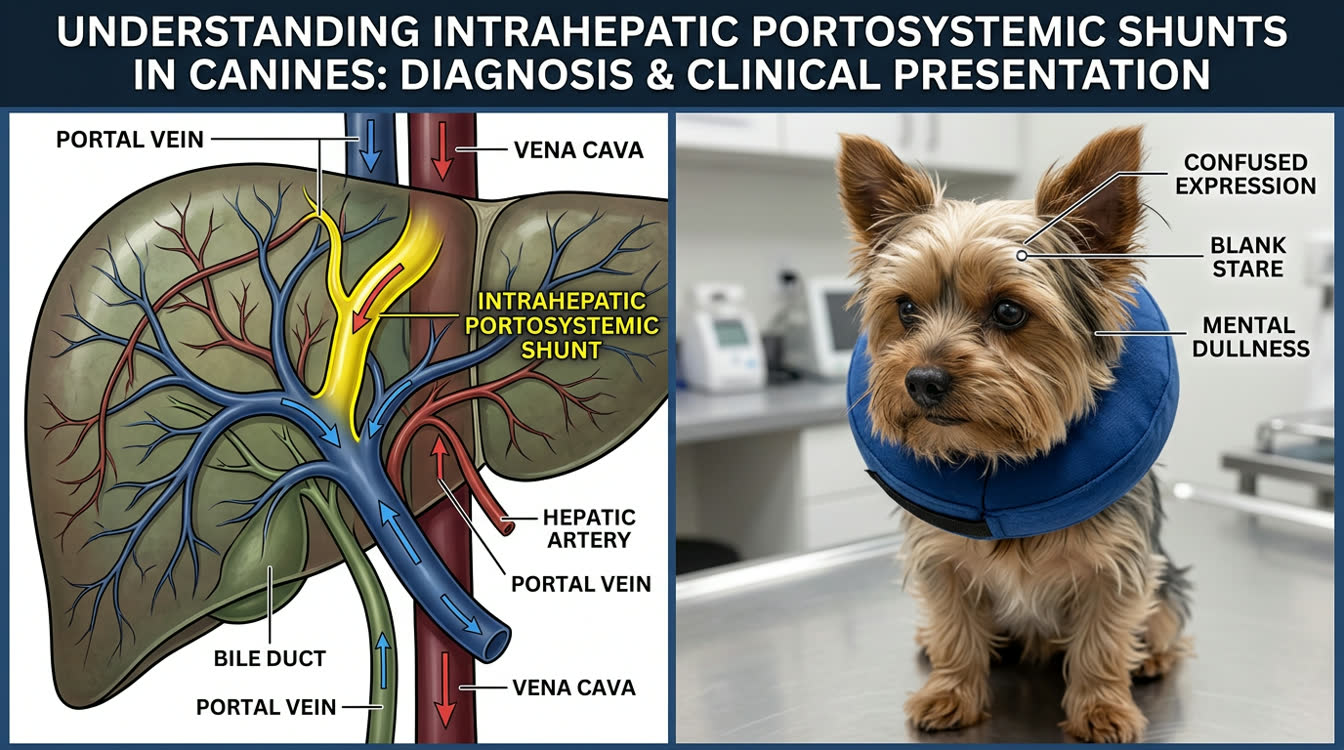
Portosystemic shunts (PSS) divert portal blood away from the liver, causing hepatic encephalopathy, urinary ammonium biurate crystals, and failure to thrive. This guide covers diagnosis, liver function tests, medical management, and surgical outcomes.
Key Takeaways
- ✓Post-prandial serum bile acids >25 µmol/L suggest PSS; CT angiography is the gold standard for surgical planning.
- ✓Ammonium biurate ("thorn-apple") crystals in urine sediment are a classic finding in dogs with PSS.
- ✓Lactulose 0.25–0.5 mL/kg PO q8–12h reduces enteric ammonia absorption and is titrated to 2–3 soft stools daily.
- ✓Phenobarbital is contraindicated in PSS — use levetiracetam 20 mg/kg q8h for seizure management.
- ✓Ameroid constrictors are the most common surgical technique for extrahepatic PSS, with 85–95% successful occlusion rates.
- ✓Post-surgical seizures occur in 5–8% of extrahepatic PSS cases within the first week; prophylactic levetiracetam is recommended.
Diagnosing PSS: Clinical Signs, Bile Acids, Ammonia and Imaging
Portosystemic shunts (PSS) are anomalous vascular connections that allow portal blood to bypass the liver and drain directly into systemic circulation. They may be congenital (single shunts) or acquired (multiple, secondary to portal hypertension from hepatic disease).
Predisposed Breeds:
- Extrahepatic PSS: Yorkshire Terrier, Maltese, Bichon Frise, Miniature Schnauzer, Pug
- Intrahepatic PSS: Irish Wolfhound, Golden Retriever, Labrador Retriever, Samoyed
Clinical Signs:
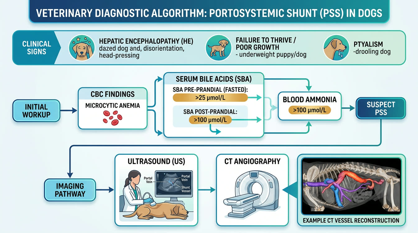
- Hepatic encephalopathy: behavioral changes, circling, head-pressing, ataxia, seizures, coma
- Failure to thrive: poor body condition, stunted growth in young animals
- Post-prandial deterioration: signs worsen 2–6 hours after protein meals
- Ptyalism (hypersalivation) — particularly common in cats with PSS
- Polyuria/polydipsia
- Urinary signs: uroliths (ammonium biurate — "thorn-apple" crystals on urine sediment), dysuria, hematuria
Diagnostic Tests:
- 12-hour fasted SBA and 2-hour post-prandial SBA
- Post-prandial SBA >25 µmol/L is highly suggestive of portovascular abnormality
- Post-prandial SBA >100 µmol/L strongly suggests PSS
- Sensitivity for PSS: ~90–95%; specificity varies with cut-off
Blood Ammonia:
- Fasting ammonia >100 µmol/L (or above lab reference range) supports hepatic encephalopathy
- Pre/post-exercise ammonia tolerance test: doubles accuracy but requires careful patient selection
- Ammonia is unstable; must be analyzed within 30 min or stored on ice
Routine Blood Work:
- Microcytic anemia (iron redistribution due to hepatic dysfunction)
- Low BUN (decreased urea synthesis)
- Hypoalbuminemia, hypoglycemia (severe cases)
- Mildly elevated ALT/ALP (variable; often normal)
- Prolonged coagulation times (factors II, VII, IX, X synthesized in liver)
Imaging:
- Abdominal ultrasound: identify shunt vessel location; microhepatica (small liver); identifies intrahepatic vs extrahepatic
- CT angiography (gold standard imaging): precisely maps shunt anatomy, diameter, and location — essential for surgical planning
- Scintigraphy (trans-splenic portal scintigraphy): quantifies shunt fraction; minimally invasive but less anatomic detail
Urinalysis:
- Ammonium biurate ("thorn-apple") crystals on sediment — highly suggestive of PSS or hepatic dysfunction
- May have proteinuria, hematuria if urolith present
Medical Management: Lactulose, Low-Protein Diet, Antibiotics and Seizure Control
Medical management is used as short-term stabilization before surgery, long-term treatment for poor surgical candidates, and for acquired PSS (portal hypertension cases where surgery is contraindicated).
Dietary Management — Foundation of Medical Therapy:
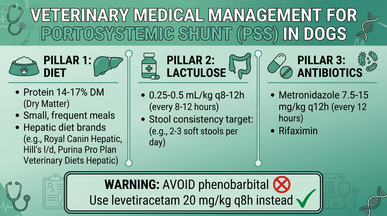
- Moderately protein-restricted, highly digestible diet
- Target: 14–17% protein DM for dogs (not severely restricted — avoid muscle wasting and impaired urea cycle)
- Preferred protein sources: dairy (casein), soy, or egg (lower ammoniagenic potential than meat)
- High-carbohydrate, highly fermentable fiber content promotes enteric ammonia trapping
- Feed multiple small meals (3–4x daily) to reduce post-prandial ammonia spikes
- Prescription hepatic diets (Royal Canin Hepatic, Hill's l/d) are appropriate
Lactulose — Enteric Ammonia Reduction:
- Mechanism: osmotic cathartic + acidification of colon (NH3 → NH4+ trapped in stool)
- Dose: 0.25–0.5 mL/kg PO q8–12h; titrate to 2–3 soft stools per day
- Use syrup formulation; avoid propylene glycol-containing products in cats
- Can be given as retention enema (diluted 3:1 with warm water) in encephalopathic animals
Antibiotics — Reduce Urease-Producing Gut Flora:
- Neomycin: 20 mg/kg PO q12h — historically used; poor systemic absorption; GI side effects
- Metronidazole: 7.5–15 mg/kg PO q12h — preferred; also has anti-inflammatory GI effects; use with caution in HE (can cause neurotoxicity at high doses)
- Rifaximin: 2.5–5 mg/kg PO q12h — minimal systemic absorption; emerging evidence in small animals
- Course: 4–8 weeks; reassess; antibiotics are adjunctive to dietary management
Seizure Management:
- Phenobarbital is AVOIDED in PSS — hepatic metabolism impaired, accumulation risk, direct hepatotoxicity
- Levetiracetam (Keppra) is preferred: 20 mg/kg PO/IV q8h — not hepatically metabolized, renal excretion
- Potassium bromide — adjunctive if needed; slow onset; use with care given renal clearance
- For acute seizure clusters: midazolam 0.1–0.2 mg/kg IN/IV is safest benzodiazepine
IV Fluids for Encephalopathic Episodes:
- 0.9% NaCl or Plasma-Lyte without lactate (lactate must be metabolized by liver)
- Supplement potassium (hypokalemia worsens encephalopathy)
- Dextrose supplementation if hypoglycemic (2.5–5% CRI)
- Avoid lactated Ringer's solution in severe hepatic failure
Surgical Correction: Ligation, Ameroid Constrictors and Outcomes
Surgical correction is the treatment of choice for congenital extrahepatic PSS in dogs. It offers the best long-term prognosis.
Surgical Options:
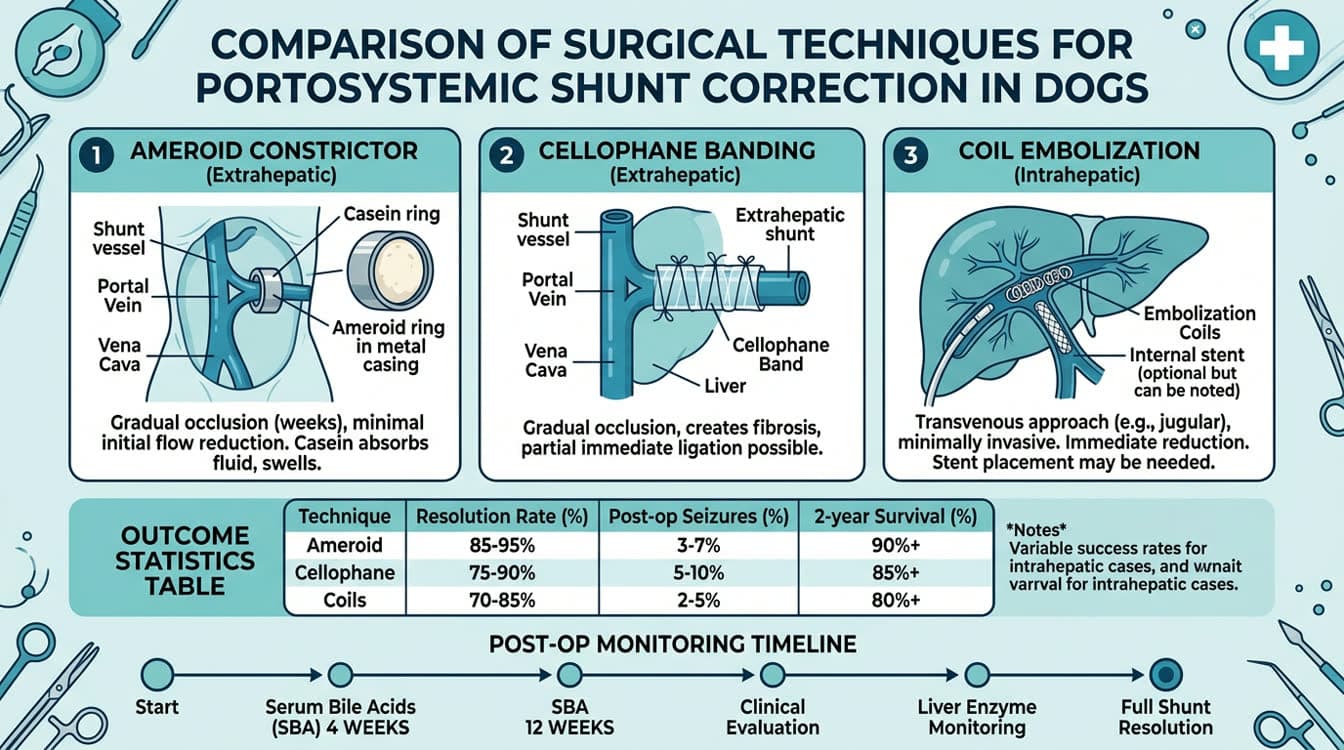
1. Ameroid Constrictor (most common for extrahepatic PSS):
- Casein ring that gradually swells over 4–8 weeks as it absorbs fluid, progressively occluding the shunt vessel
- Gradual occlusion reduces risk of acute portal hypertension
- Complete occlusion confirmed at 4–8 weeks; partial occlusion in ~15–20% of cases
2. Cellophane Band Attenuation:
- Cellophane band placed around the shunt vessel; fibrous reaction gradually occludes over weeks
- Attrition rate similar to ameroid
3. Intravascular Coil Embolization (intrahepatic PSS):
- Interventional radiologic technique; coils placed within the shunt vessel via transjugular or transabdominal approach
- Preferred for intrahepatic PSS (right-divisional or left-divisional)
- Requires specialist center with interventional radiology capability
4. Acute Ligation:
- Sudden occlusion carries high risk of fatal portal hypertension
- Only used in dogs with good portal tolerance demonstrated intraoperatively (portal pressure <20 cmH2O after test occlusion)
Surgical Outcomes:
| Metric | Extrahepatic PSS | Intrahepatic PSS |
|---|---|---|
| Successful occlusion | 85–95% | 70–85% |
| Resolution of clinical signs | 75–85% | 65–80% |
| Post-op seizure rate | 5–8% | 8–15% |
| Long-term survival (2 yr) | 85–90% | 75–85% |
| Surgical mortality | 2–5% | 5–10% |
Post-Surgical Management:
- Continue medical management (diet, lactulose) for 4–8 weeks post-surgery until adequate occlusion confirmed
- Post-op SBA at 4 and 12 weeks — if still markedly elevated, assess for residual shunting
- Monitor for: acute portal hypertension (abdominal pain, hemorrhagic diarrhea), seizures (2–7 days post-op), hypoglycemia
- Levetiracetam 20 mg/kg PO q8h for first 2 weeks post-surgery to reduce seizure risk
When Surgery Is Contraindicated:
- Acquired PSS from portal hypertension (hepatic cirrhosis): multiple small vessels — surgical closure worsens portal hypertension
- Severe hepatic dysfunction (albumin <1.5 g/dL, prolonged PT >2x normal): high anesthetic/surgical risk
- These patients are managed long-term medically with diet, lactulose, and antibiotics
Frequently Asked Questions
What is the most reliable test to diagnose a portosystemic shunt in a dog?
Post-prandial serum bile acids (SBA) are the gold standard function test — >25 µmol/L is suggestive and >100 µmol/L is strongly indicative of PSS. CT angiography is the gold standard for anatomical diagnosis and surgical planning.
Why is phenobarbital avoided in dogs with PSS?
Phenobarbital is extensively metabolized by the liver and is directly hepatotoxic. In dogs with a PSS, hepatic dysfunction leads to drug accumulation, increased neurotoxicity risk, and worsening liver disease. Levetiracetam (Keppra) is the preferred anticonvulsant because it is renally eliminated.
Can a dog with PSS survive without surgery?
Some dogs with mild disease can live for years with medical management (diet, lactulose, antibiotics). However, surgery offers significantly better long-term outcomes (85–90% 2-year survival) compared to lifelong medical management for extrahepatic PSS. Medical management is primarily for stabilization before surgery or for inoperable cases.
What does an ammonium biurate crystal in urine mean?
Ammonium biurate ("thorn-apple") crystals in urine sediment indicate hyperammonemia, which is strongly associated with PSS or severe liver disease. They can also form uroliths causing dysuria and hematuria. Urate crystalluria warrants evaluation with serum bile acids.
What breeds are most at risk for portosystemic shunts?
Yorkshire Terriers and Maltese are most commonly affected by extrahepatic PSS. Large breeds including Irish Wolfhounds, Golden Retrievers, and Labrador Retrievers are predisposed to intrahepatic PSS. Genetic inheritance is suspected in many affected breeds.
References
- Greenhalgh SN, et al. (2014). Long-term survival and quality of life in dogs with clinical signs associated with a congenital portosystemic shunt after surgical or medical treatment. J Am Vet Med Assoc. 245(5):527–533.
- Weisse C, et al. (2014). Percutaneous transvenous coil embolization of hepatic intravascular portosystemic shunts. J Vet Intern Med. 28(2):362–370.
- Tivers MS, et al. (2014). Post-operative neurological signs in dogs following surgical management of single congenital portosystemic shunts. Vet Rec. 174(12):297–302.