Quick Answer
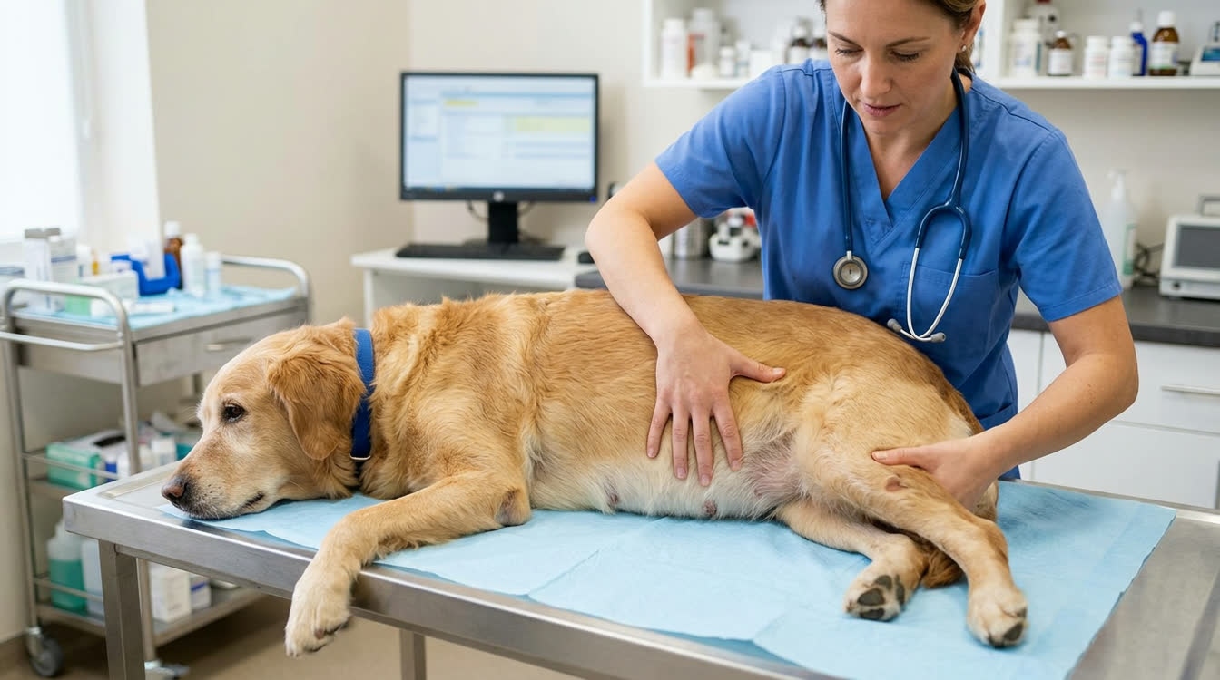
Protein-losing enteropathy (PLE) causes protein loss through the GI tract resulting in hypoalbuminemia, edema, and ascites. This guide covers causes including lymphangiectasia and IBD, diagnostic workup, low-fat dietary management, and immunosuppressive therapy.
Key Takeaways
- ✓PLE causes panhypoproteinemia (both albumin AND globulins low) -- key distinction from liver disease
- ✓Ultra-low-fat diet (<10-12% fat DM) is essential for lymphangiectasia
- ✓fecal alpha-1-protease inhibitor (fA1PI) is a sensitive marker to confirm intestinal protein loss
- ✓Yorkshire Terriers and Soft Coated Wheaten Terriers have breed-specific PLE predispositions
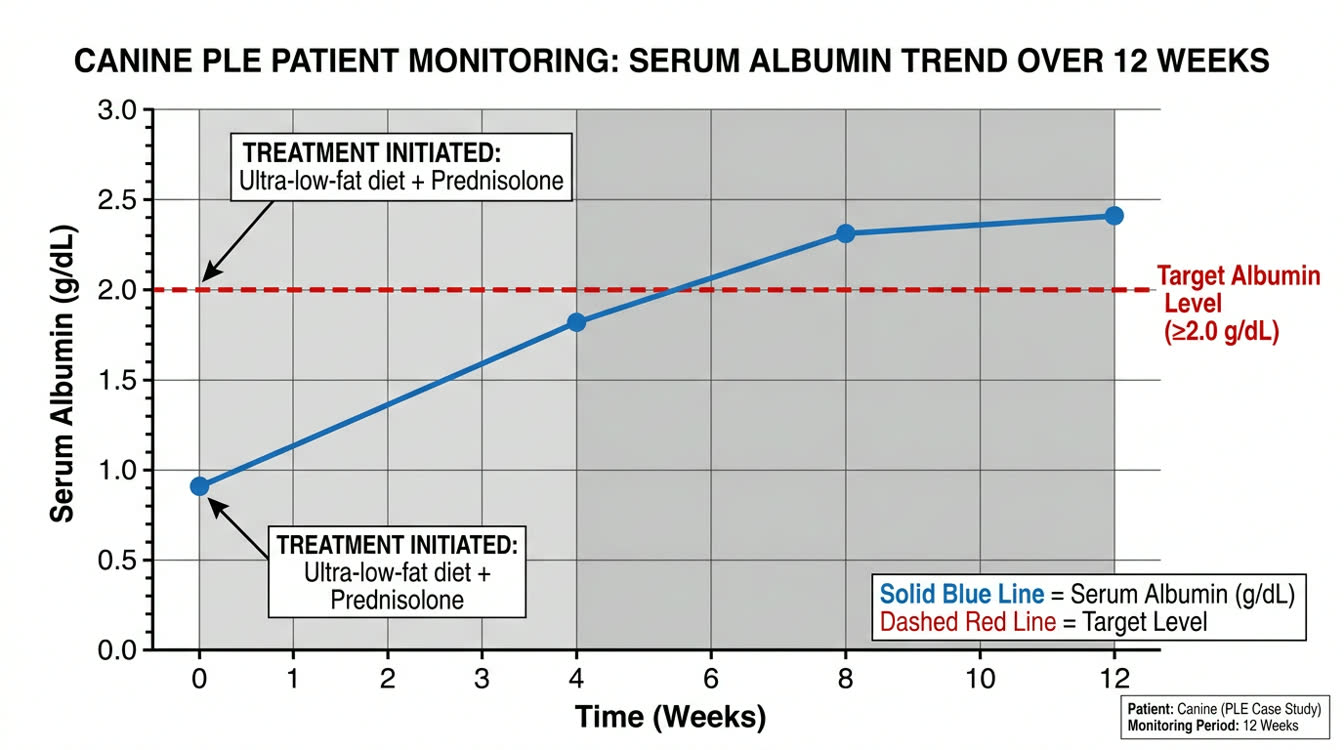
- ✓Monitor albumin every 2-4 weeks; target >2.0 g/dL to prevent edema and ascites
Causes and Diagnosis of PLE
What Is PLE? PLE is a syndrome of excessive protein loss through the GI tract. Hypoalbuminemia reduces oncotic pressure causing edema, ascites, and pleural effusion.
Major Causes
| Cause | Key Features |
|---|---|
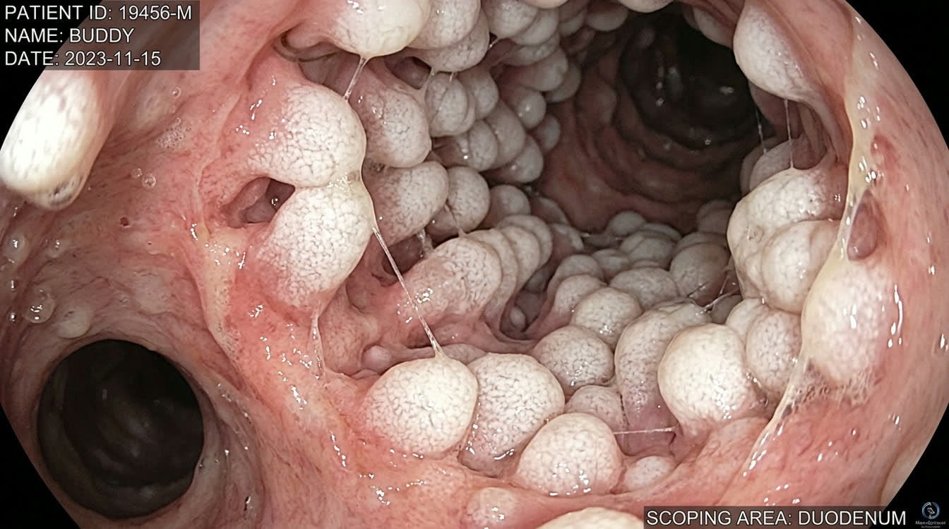
| Intestinal lymphangiectasia | Dilated lacteals on biopsy; Yorkshire Terrier predisposed |
| IBD (lymphoplasmacytic, eosinophilic) | Inflammatory infiltrate on biopsy |
| Intestinal lymphoma (T-cell) | IHC and PARR testing |
| Fungal (Pythium, Histoplasma) | Titers; biopsy with organisms |
Breed Predispositions
- Yorkshire Terrier: primary lymphangiectasia
- Soft Coated Wheaten Terrier: concurrent protein-losing nephropathy (PLN)
- Norwegian Lundehund, Rottweiler, Irish Setter: overrepresented
Clinical Signs
- Hypoalbuminemia (albumin <2.0 g/dL)
- Weight loss, chronic diarrhea
- Ascites, pitting edema (ventral)
- Pleural effusion in severe cases
Diagnostic Workup 1. Serum albumin: <2.0 g/dL suggests PLE; <1.5 g/dL severe 2. Panhypoproteinemia (both albumin AND globulins low) -- vs liver disease where globulins elevated 3. Urinalysis + UPC ratio: rule out PLN (concurrent in Wheaten Terriers) 4. Fecal alpha-1-protease inhibitor (fA1PI): confirms intestinal protein loss; Texas A&M GI Lab 5. Abdominal ultrasound: layering, lymphadenopathy, hyperechoic mucosal striations (dilated lacteals) 6. Endoscopy/biopsy: gold standard
Ultra-Low-Fat Dietary Management
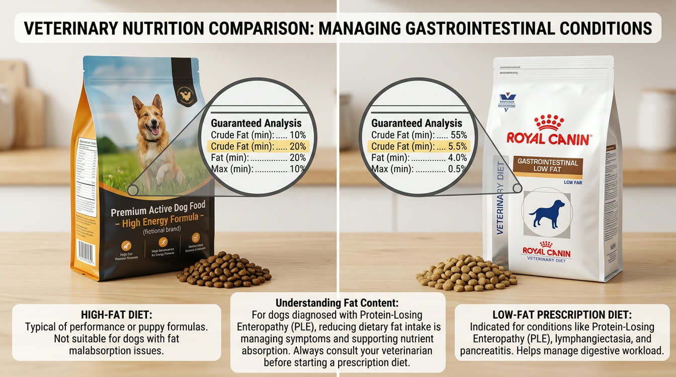
Why Low-Fat Diet Is Critical Dietary fat stimulates lymphatic flow. In lymphangiectasia, high-fat meals increase lactal pressure and protein leakage. Ultra-low-fat diet (<10-12% fat DM) is essential.
Diet Options
| Diet | Approximate Fat (DM) |
|---|---|
| Royal Canin Gastrointestinal Low Fat | ~9% |
| Hill's i/d | ~14% |
| Home-cooked: boiled chicken breast + white rice | ~5-8% |
Nutritional Support
- 25% human serum albumin 2 mL/kg IV over 6-8 hours: pretreat with diphenhydramine 2 mg/kg IV
- Fresh frozen plasma 6-10 mL/kg: temporary albumin support
- Fat-soluble vitamins (A, D, E, K) often deficient -- supplement orally
- Cobalamin supplementation if deficient (check serum B12)
Immunosuppression and Albumin Monitoring
Immunosuppression for IBD-Associated PLE
First-Line: Prednisolone
- 2 mg/kg PO q24h (immunosuppressive dose) x 4-6 weeks
- Taper: reduce by 25-50% every 2-4 weeks based on response
- Maintenance: 0.5-1 mg/kg q48h or lowest effective dose
Second-Line: Azathioprine (dogs only)
- 2 mg/kg PO q24h x 2-4 weeks, then 1-2 mg/kg q48h
- CBC q2-4 weeks (myelosuppression); liver enzymes q3 months
Alternative: Chlorambucil
- 2-6 mg/m2 PO q24-48h: for T-cell lymphoma IBD or refractory cases
Monitoring
| Parameter | Frequency | Target |
|---|---|---|
| Serum albumin | q2-4 weeks initially | >2.0 g/dL |
| Body weight | Weekly | Stable or gaining |
| CBC (if on azathioprine) | q2-4 weeks | Normal WBC/platelets |
Prognosis
- Lymphangiectasia (primary): fair to good with lifelong low-fat diet
- IBD: variable; good if responsive to corticosteroids
- T-cell lymphoma: guarded (median 4-6 months)
Related Articles:
- [Megaesophagus in Dogs](/articles/megaesophagus-dogs)
- [Exocrine Pancreatic Insufficiency in Dogs](/articles/exocrine-pancreatic-insufficiency-dogs)
Frequently Asked Questions
What causes PLE in dogs?
The three most common causes are intestinal lymphangiectasia, IBD, and intestinal lymphoma. Yorkshire Terriers are particularly predisposed to lymphangiectasia. Less common: fungal infections (Pythium, Histoplasma), intestinal adenocarcinoma, and severe GI ulceration.
How low does my dog's diet fat need to be for PLE?
For lymphangiectasia, <10-12% fat on dry matter basis. Royal Canin Gastrointestinal Low Fat (~9% DM) or home-cooked boiled chicken breast + white rice (~5-8% DM) work well. Verify fat content with your veterinarian.
Why is albumin important to monitor?
Albumin maintains oncotic pressure. Below 2.0 g/dL it causes ascites, edema, and pleural effusion. Below 1.5 g/dL risks hypovolemia and hypocalcemia. Monitor every 2-4 weeks initially to guide treatment adjustments.
When is immunosuppression needed?
When intestinal biopsy confirms IBD as the underlying cause, prednisolone 2 mg/kg PO q24h is indicated. Dogs with lymphangiectasia may not need it unless concurrent IBD is present. Always biopsy before starting immunosuppression.
References
- Dossin O, Lavoue R. Protein-losing enteropathies in dogs. Vet Clin North Am Small Anim Pract. 2011;41(2):399-418.
- Willard MD. Protein-losing enteropathies in dogs. In: Ettinger SJ, Feldman EC, eds. Textbook of Veterinary Internal Medicine. 8th ed. Elsevier; 2017.