Quick Answer
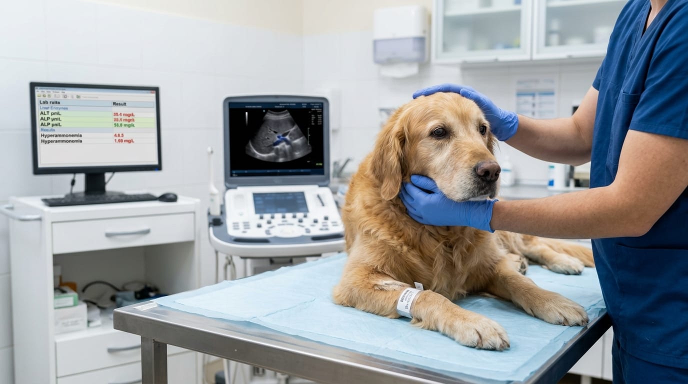
Hepatic encephalopathy (HE) is a neurological syndrome caused by liver failure or portosystemic shunting. This guide covers pathophysiology, clinical grading, acute management with lactulose enemas and levetiracetam, and triggers to address.
Key Takeaways
- ✓Hepatic encephalopathy is graded I–IV based on clinical signs; Grade III–IV (stupor/coma) requires emergency hospitalization.
- ✓Ammonia is a primary but imperfect marker — clinical grade and response to treatment are more informative than a single ammonia value.
- ✓Lactulose retention enemas (1:3 dilution, 5–10 mL/kg, q4–6h) are the most rapid-acting intervention for acute HE.
- ✓Phenobarbital and lactated Ringer's solution are contraindicated in hepatic failure — use levetiracetam and Plasma-Lyte A instead.
- ✓Moderate (not zero) protein restriction with dairy, soy, or egg protein prevents muscle catabolism while reducing ammonia generation.
- ✓Identify and correct precipitating triggers: hypokalemia, constipation, GI hemorrhage, infection, and high-protein meals.
Pathophysiology and Clinical Grading of Hepatic Encephalopathy
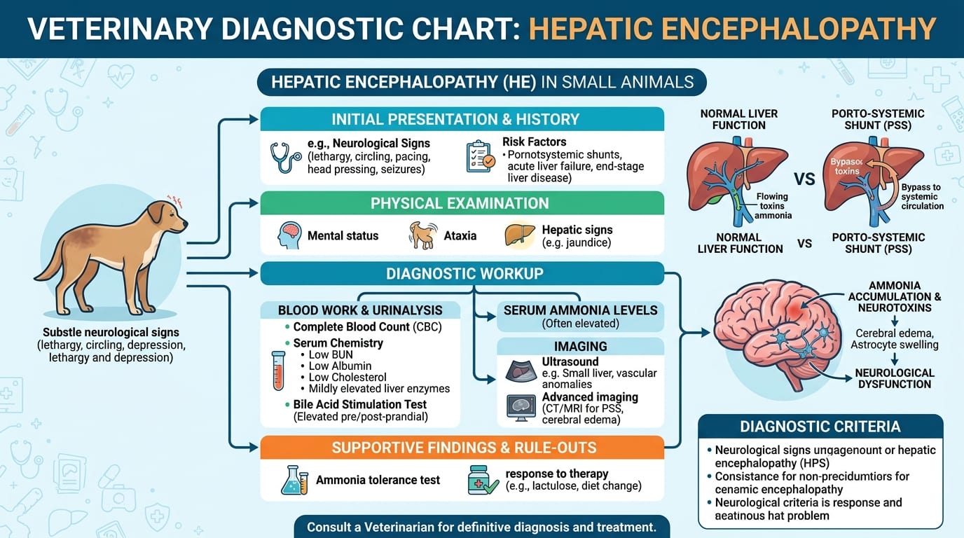
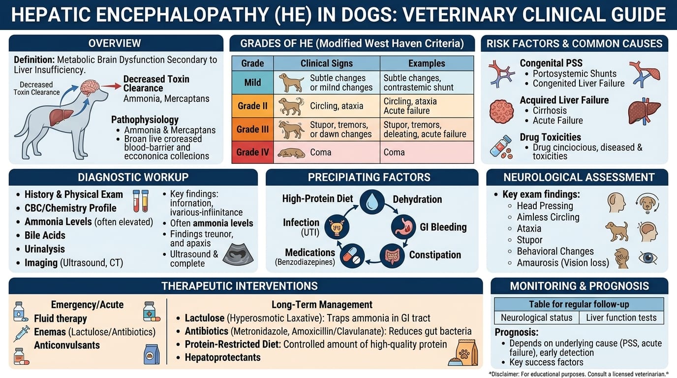
Hepatic encephalopathy (HE) is a metabolic neurological syndrome arising from the failure of the liver to clear neurotoxic substances from portal blood. In small animals, the primary causes are portosystemic shunts (PSS) and severe hepatic parenchymal failure.
Primary Neurotoxins in HE:
Ammonia (NH3):
- Produced in the colon by urease-producing bacteria (Escherichia, Proteus, Urease-positive organisms) and from amino acid catabolism
- Normally converted to urea in the liver (urea cycle)
- Excess ammonia crosses the blood-brain barrier and causes astrocyte swelling, glutamate-glutamine imbalance, and mitochondrial dysfunction
- Blood ammonia does not correlate perfectly with clinical signs — other toxins also contribute
Other Contributing Factors:
- GABA (gamma-aminobutyric acid): increased GABAergic neurotransmission (false neurotransmitters, benzodiazepine-like compounds from gut flora)
- Aromatic amino acids: tyrosine, phenylalanine compete with branched-chain amino acids for BBB transport, generating false neurotransmitters (octopamine)
- Manganese: accumulates in PSS patients; deposits in basal ganglia
- Systemic inflammation: amplifies encephalopathy severity
Precipitating Triggers of HE Episodes:
- High-protein meal
- GI hemorrhage (digested blood = protein load)
- Constipation (prolonged bacterial ammonia production)
- Hypokalemia (promotes renal ammonia generation)
- Alkalosis (increases uncharged NH3 BBB permeability)
- Infection/sepsis
- Sedatives, opioids (CNS depression synergism)
- Azotemia (increased urea → ammonia conversion)
- Dehydration
Clinical Grading Scale (modified from human ISHEN):
| Grade | Clinical Signs |
|---|---|
| 0 | Subclinical — normal exam; cognitive deficits on testing only |
| I | Mild confusion, personality change, mild ataxia, hypersalivation |
| II | Disorientation, circling, head-pressing, blindness, drowsiness |
| III | Stupor — semi-conscious, responds only to pain |
| IV | Coma — no response to stimuli; seizures possible |
Key Physical Exam Findings:
- Microhepatica (PSS): small liver on palpation or imaging
- Icterus: may suggest severe parenchymal failure rather than PSS
- Ascites: portal hypertension component
- Ammonium biurate crystalluria: urine sediment
Acute HE Management: Lactulose Enemas, Fluids and Seizure Control
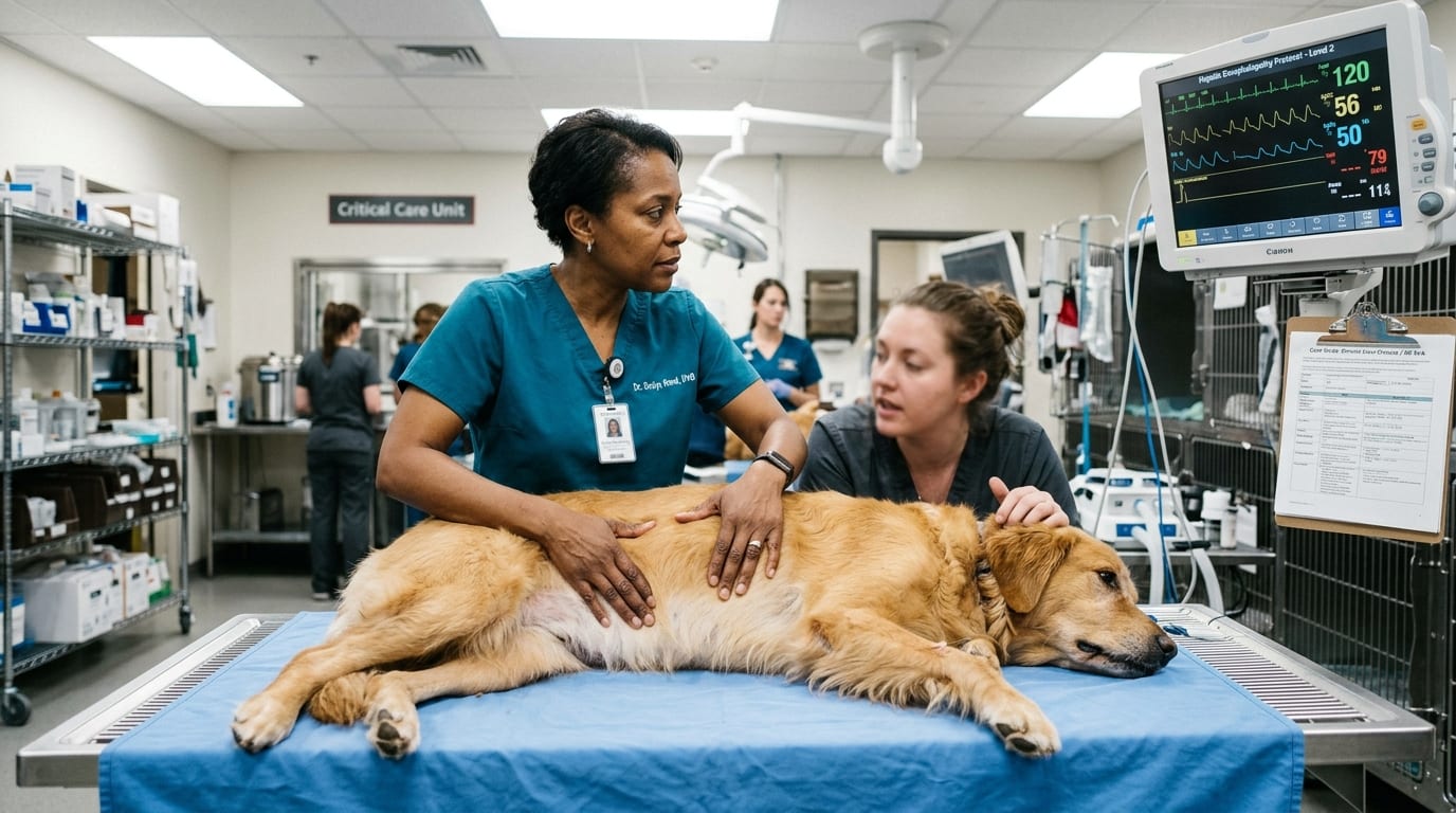
Acute Grade III–IV HE is a veterinary emergency requiring aggressive intervention to reduce blood ammonia and neurological toxin load.
Immediate Priorities: 1. Maintain airway — intubation in Grade IV coma patients 2. IV access — start fluids immediately 3. Check blood glucose — HE-associated hypoglycemia is common and life-threatening 4. Identify and treat precipitating trigger
IV Fluid Therapy:
- 0.9% NaCl or Plasma-Lyte A — do NOT use lactated Ringer's in severe hepatic failure (impaired lactate metabolism)
- Supplement potassium: target K+ 3.5–5.0 mEq/L (hypokalemia worsens HE)
- 2.5–5% dextrose CRI if hypoglycemic (target BG 80–140 mg/dL)
- Avoid excessive fluid rates — risk of cerebral edema in advanced HE
Lactulose Retention Enema (most impactful acute intervention):
- Dilute lactulose 1:3 (1 part lactulose : 3 parts warm water)
- Volume: 5–10 mL/kg per enema (warm, not hot)
- Retained for 15–20 minutes if possible; gentle massage of abdomen
- Repeat q4–6h until fecal ammonia reduction achieved (soft stool, patient improving)
- Mechanism: colonic acidification traps NH4+ and osmotic purging reduces bacterial burden
Oral Lactulose (when eating or NG tube):
- 0.25–0.5 mL/kg PO q8h; titrate to 2–3 soft stools daily
Antibiotic Therapy:
- Metronidazole: 7.5–15 mg/kg PO q12h — reduces urease-producing bacteria; also has anti-inflammatory GI effect
- Rifaximin: 2.5–5 mg/kg PO q12h — emerging; non-absorbable, minimal systemic effects
Seizure Management:
- Levetiracetam (drug of choice): 20 mg/kg IV/PO q8h — no hepatic metabolism
- Diazepam: Avoid in chronic HE (may worsen encephalopathy via GABA pathway); acceptable for acute cluster seizures at 0.5 mg/kg IV once
- Phenobarbital: CONTRAINDICATED — hepatotoxic, accumulates in hepatic failure
- Propofol CRI: 0.1–0.4 mg/kg/min IV for refractory status — metabolized by extrahepatic tissues (not solely hepatic)
Nutritional Support:
- NPO initially in Grade III–IV; transition to enteral feeding when patient is Grade I–II
- Nasogastric or nasoesophageal tube feeding with hepatic diet
- Target: caloric intake 70–80 kcal/kg/day; moderate protein (avoid zero-protein diets — muscle catabolism worsens ammonia)
Long-Term Management, Monitoring and Trigger Avoidance
After stabilizing an acute HE episode, long-term management focuses on minimizing ammonia load, addressing the underlying cause, and preventing recurrence.
Dietary Management:
- Moderately protein-restricted diet: 14–17% protein DM (dogs); avoid severe restriction — leads to catabolism
- Preferred protein sources: dairy, soy, egg (lower ammoniagenic than red meat)
- Royal Canin Hepatic, Hill's l/d, Purina NF or HP: all appropriate depending on underlying disease
- Feed 3–4 small meals per day to blunt post-prandial ammonia spikes
- Zinc supplementation (zinc acetate 2–5 mg/kg/day PO): reduces gut ammonia production; supports urea cycle enzymes
Trigger Avoidance Protocol:
| Trigger | Prevention Strategy |
|---|---|
| High-protein meals | Divide daily ration into 3–4 small meals |
| GI hemorrhage | Treat ulcers with sucralfate + famotidine |
| Constipation | Maintain lactulose at stool-softening dose |
| Hypokalemia | Supplement K+ in diet; check electrolytes q3 months |
| Infections | Treat promptly; avoid bacteremia |
| Sedatives/opioids | Minimize; choose drugs with extrahepatic metabolism |
Monitoring Schedule:
- Baseline: SBA, ammonia, CBC, chemistry, UA
- 1 month post-treatment: ammonia, SBA, clinical assessment
- Every 3 months: SBA, chemistry, electrolytes (K+)
- 6–12 months: echocardiogram/CT (if surgical PSS follow-up)
Drugs to Avoid in HE:
- Phenobarbital, benzodiazepines (chronic use)
- Tetracyclines (hepatotoxic at higher doses)
- Sulfonamides (hepatotoxic in sensitive individuals)
- Acetaminophen (dogs with hepatic disease)
- NSAIDs (GI hemorrhage risk worsens ammonia load)
- Azathioprine (some hepatic metabolism; monitor carefully)
When to Refer:
- Refractory Grade III–IV HE despite maximum medical management
- Suspected PSS for surgical evaluation (CT angiography)
- Suspected underlying hepatic neoplasia or inflammatory hepatopathy requiring biopsy
- Persistent coagulopathy requiring specialty management
Frequently Asked Questions
Does blood ammonia level correlate with hepatic encephalopathy severity?
Not perfectly. While hyperammonemia contributes to HE, blood ammonia levels do not reliably predict clinical grade. Some dogs with markedly elevated ammonia show mild signs, while others with borderline ammonia levels are severely encephalopathic. Clinical grading and response to treatment are more meaningful than a single ammonia value.
What is the fastest way to reduce ammonia in an encephalopathic dog?
Lactulose retention enemas (diluted 1:3 with warm water, 5–10 mL/kg, retained 15–20 minutes, q4–6h) are the most rapidly effective intervention for reducing enteric ammonia load in acute HE. IV fluid therapy to correct hypokalemia and hypoglycemia is equally important.
Can diazepam be used for seizures in a dog with hepatic encephalopathy?
Diazepam should be used very cautiously in HE. Chronic benzodiazepine use may worsen encephalopathy by augmenting GABAergic pathways. Levetiracetam is the drug of choice for seizures in HE. A single dose of diazepam for an acute cluster seizure is acceptable, but repeated or continuous use is avoided.
Should I feed a dog with hepatic encephalopathy a zero-protein diet?
No. Severe protein restriction (zero protein) causes muscle catabolism, which generates ammonia from endogenous protein breakdown — counterproductive. The goal is moderate protein restriction (14–17% DM) using low-ammoniagenic protein sources like dairy, soy, or egg, fed in multiple small meals.
What role does zinc play in hepatic encephalopathy management?
Zinc acts as a urea cycle cofactor and reduces gut ammonia production by inhibiting urease activity. Zinc acetate 2–5 mg/kg/day PO is an adjunctive treatment for HE, particularly in PSS patients. Zinc deficiency is common in hepatic disease and contributes to HE severity.
References
- Lidbury JA, et al. (2015). Hepatic encephalopathy in dogs and cats. J Vet Emerg Crit Care. 25(4):471–487.
- Center SA. (2007). Interpretation of liver enzymes. Vet Clin North Am Small Anim Pract. 37(2):297–333.
- Rothuizen J, et al. (2006). WSAVA Standards for Clinical and Histological Diagnosis of Canine and Feline Liver Diseases. Elsevier.