Quick Answer
Hepatic lipidosis is the most common feline hepatopathy and results from prolonged anorexia and hepatic fat accumulation. This guide covers the pathophysiology, diagnostic criteria, enteral feeding tube selection and placement, caloric targets, refeeding syndrome prevention, and prognosis with aggressive nutritional support.
Key Takeaways
- ✓Hepatic lipidosis develops within 2-10 days of anorexia in cats, especially obese cats; any cat not eating for 48-72 hours needs urgent evaluation.
- ✓Diagnosis is confirmed by hepatic FNA cytology showing lipid-vacuolated hepatocytes; ultrasound shows diffuse hyperechoic liver.
- ✓Esophagostomy tube placement is the gold standard for enteral nutrition; begin at 25% RER and increase to 100% over 4-5 days.
- ✓Vitamin K1 (0.5-1.5 mg/kg SQ) must be given before tube placement or any invasive procedure due to hepatic coagulopathy.
- ✓Refeeding syndrome prevention: monitor phosphorus every 24-48 hours; supplement thiamine 50-100 mg daily; increase calories gradually.
- ✓Prognosis with aggressive enteral nutritional support: 60-85% survival; cats with primary idiopathic HL have better outcomes than secondary HL.
Pathophysiology, Clinical Presentation and Diagnostic Criteria
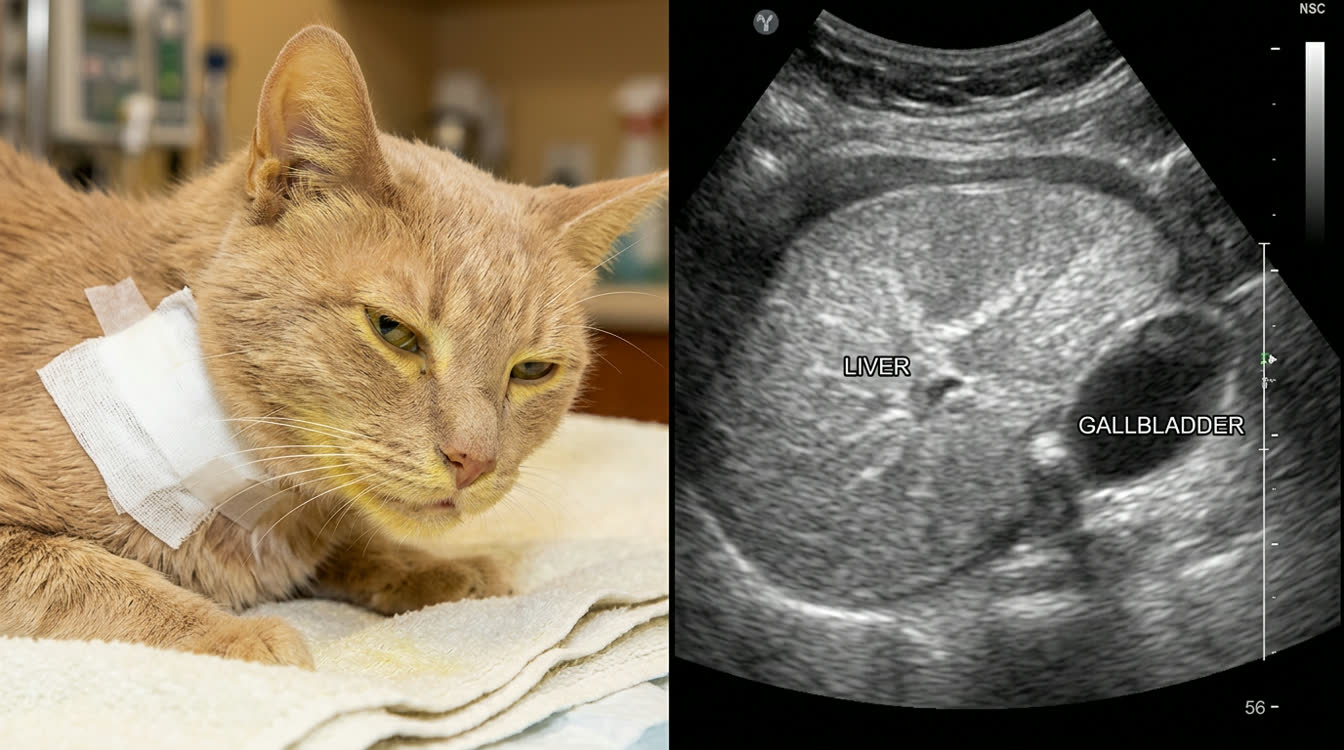
Hepatic lipidosis (HL) is the most common hepatobiliary disease in cats and one of the most common causes of jaundice. It can develop rapidly — within 2-10 days of anorexia in obese cats.
Pathophysiology
- Progressive hepatocyte dysfunction
- Intrahepatic cholestasis
- Obstructive jaundice
- Coagulopathy (reduced hepatic production of clotting factors)
Risk Factors and Triggering Events
- Obesity is the primary risk factor (obese cats mobilize more adipose)
- Any cause of anorexia lasting > 2-3 days can trigger HL: stress, illness (URI, pancreatitis, IBD), dietary change, environmental change
- Primary idiopathic HL occurs in cats that stop eating for no identifiable reason
Clinical Signs
- Profound weakness, obtundation
- Anorexia (often weeks of partial or complete anorexia by presentation)
- Icterus (jaundice): scleral, mucosal, skin
- Ptyalism (drooling) — from hepatic encephalopathy
- Muscle wasting, ventroflexion of the neck (hypokalemia)
- Vomiting, hepatomegaly
Diagnostic Criteria
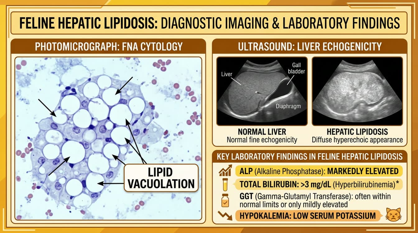
- Chemistry: elevated ALT, ALP (ALP markedly elevated — ALP induction by lipid accumulation is unique to HL); elevated bilirubin (total > 2-3 mg/dL in HL)
- Urinalysis: bilirubinuria
- Abdominal ultrasound: diffuse hepatomegaly with hyperechoic (bright) liver parenchyma
- Hepatic cytology (fine needle aspirate): vacuolated hepatocytes with intracellular lipid droplets (lipid vacuolation) — confirms diagnosis
- PT/aPTT: often prolonged (vitamin K-responsive coagulopathy); administer vitamin K1 before invasive procedures
- Electrolytes: hypokalemia (from anorexia, vomiting); hypophosphatemia (refeeding risk)
Esophagostomy Tube Placement, Caloric Targets and Feeding Protocols
Why Enteral Nutrition is the Cornerstone of HL Treatment Force-feeding cats by hand delivers inadequate calories and is stressful, which worsens hepatic lipidosis. Tube feeding restores anabolic state, halts lipid mobilization, and allows the liver to recover.
Feeding Tube Options
| Tube Type | Placement | Tube Size | Duration | Notes |
|---|---|---|---|---|
| Esophagostomy (E-tube) | Surgical, lateral cervical | 10-14 Fr | Weeks-months | Preferred for HL; tolerates thick diets |
| Nasogastric (NG) | Non-surgical | 5-8 Fr | 3-7 days | Short-term only; liquid diets only |
| Gastrostomy (PEG) | Endoscopic or surgical | 14-22 Fr | Months | Preferred if long-term needed post-recovery |
| Jejunostomy | Surgical | 5-8 Fr | Short-term | If vomiting prevents gastric feeding |
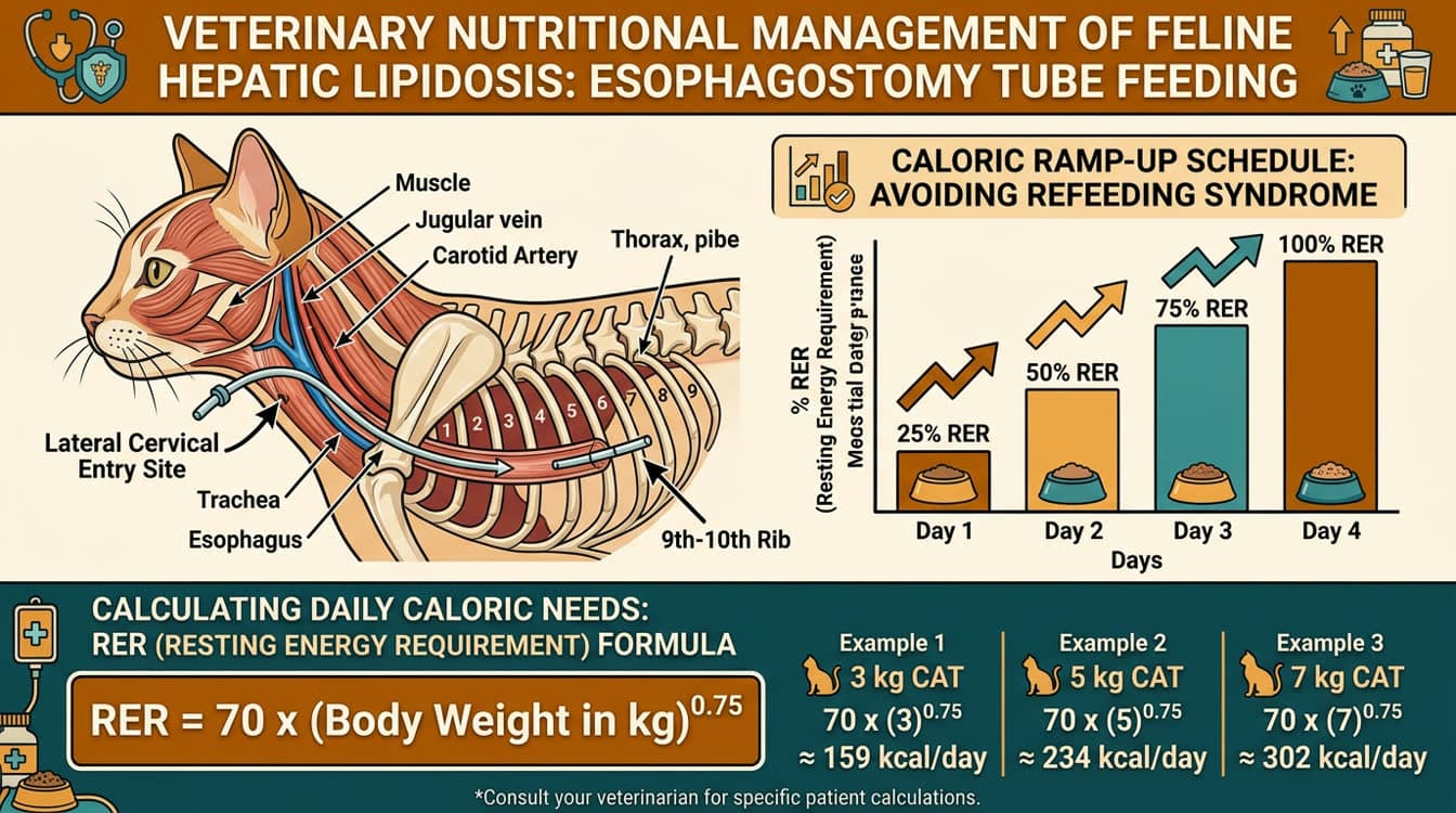
Esophagostomy Tube Placement Technique 1. Anesthesia induction (propofol 2-4 mg/kg IV; mask maintenance with isoflurane) 2. Position cat in right lateral recumbency; clip and prep left lateral cervical region 3. Insert finger (or Carmalt forceps) down esophagus to mid-cervical level; tent skin outward 4. Make stab incision over tented area; pass tube into esophagus distally to distal third 5. Anchor with Chinese finger-trap suture pattern at insertion site 6. Verify position via lateral radiograph (tube tip at 9th-10th rib level, within esophagus) 7. Protect with light neck bandage
Caloric Targets and Reintroduction Schedule
- Resting Energy Requirement (RER): RER (kcal/day) = 70 x body weight (kg)^0.75
- Target: 100% RER within 3-5 days; begin at 25-33% RER on day 1 to avoid refeeding syndrome
- Day 1: 25% RER; Day 2: 50% RER; Day 3: 75% RER; Day 4+: 100% RER
- Divide into 4-6 meals per day; flush tube with 5-10 mL warm water before and after each feeding
Diet Formulation
- High-protein, moderate-fat commercial recovery diet (Hill's a/d, Royal Canin Recovery, Purina CN)
- Blend to smooth consistency; aim for 1 kcal/mL concentration
- Do NOT use low-protein diets initially (cats require protein for hepatic recovery)
- Add taurine supplement if not already present in diet
Refeeding Syndrome Prevention, Supplements and Prognosis
Refeeding Syndrome Rapid reintroduction of nutrition can cause dangerous shifts in phosphorus, potassium, and magnesium as anabolic metabolism resumes, potentially causing cardiac arrhythmias, hemolytic anemia (hypophosphatemia), and neuromuscular weakness.
Prevention Protocol
- Start at 25% RER; increase gradually over 4-5 days
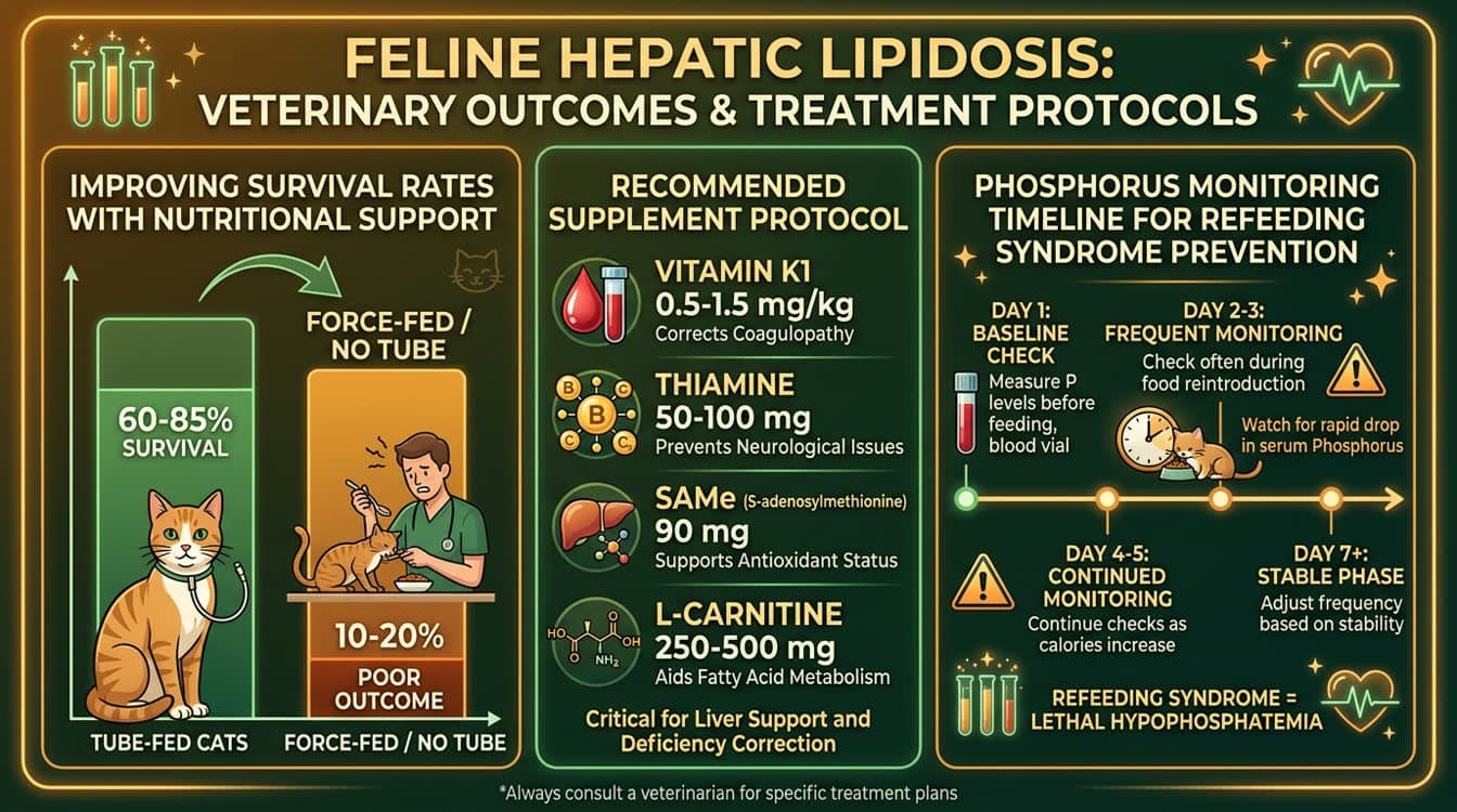
- Monitor phosphorus every 24-48 hours; target > 2.5 mg/dL
- If phosphorus < 1.5 mg/dL: potassium phosphate supplementation IV (0.01-0.03 mmol/kg/h)
- Monitor potassium daily; supplement KCl IV if < 3.0 mEq/L
- Thiamine supplementation: 50-100 mg IM or PO q24h (required — cats have high thiamine requirements and depletion worsens hepatic encephalopathy)
Key Supplements in HL Management
- Vitamin K1: 0.5-1.5 mg/kg SQ q12-24h x 3 doses before any invasive procedures (tube placement, biopsy); corrects vitamin K-responsive coagulopathy
- Thiamine (Vitamin B1): 50-100 mg IM daily; prevents Wernicke-like encephalopathy
- L-carnitine: 250-500 mg PO daily; supports fatty acid beta-oxidation in hepatocytes
- SAMe (S-adenosylmethionine): 90 mg PO q24h on empty stomach (enteric coated); hepatoprotective; supports glutathione synthesis
- Ursodeoxycholic acid (UDCA): 10-15 mg/kg PO q24h with food; choleretic, hepatoprotective; use after coagulopathy corrected
- Potassium: supplementation as needed in fluids; correct before feeding
Prognosis
- With early aggressive enteral nutritional support: survival rate 60-85%
- Primary (idiopathic) HL: better prognosis than secondary HL (underlying IBD, pancreatitis, neoplasia)
- Poor prognostic indicators: severe hepatic encephalopathy, PT/aPTT > 3x normal, bilirubin > 15 mg/dL, concurrent pancreatitis or triaditis
- Recovery timeline: most cats begin voluntarily eating within 3-6 weeks of tube feeding initiation
- Tube can be removed once cat voluntarily eats 100% of caloric needs for 3 consecutive days
Preventing Recurrence
- Maintain healthy body weight in recovered cats (obesity is primary risk factor)
- Never withhold food from a cat for more than 24-36 hours; even brief anorexia is risky
- Monitor for early signs of anorexia in cats with chronic diseases (IBD, CKD, hyperthyroidism)
Frequently Asked Questions
How quickly can hepatic lipidosis develop in a cat?
Hepatic lipidosis can develop within 2-10 days of complete anorexia, particularly in obese cats. Even partial anorexia over 1-2 weeks can trigger significant hepatic lipid accumulation. This is why any cat that has not eaten for more than 48-72 hours requires urgent veterinary evaluation.
Why is an esophagostomy tube preferred over a nasogastric tube for hepatic lipidosis?
Esophagostomy tubes are preferred because they allow feeding of thick, calorie-dense recovery diets at appropriate volumes. They are tolerated well long-term (weeks to months) and do not interfere with normal nasal function. Nasogastric tubes require liquid diets only, can only be maintained short-term (3-7 days), and are often removed by cats.
What is the caloric target for tube-feeding a cat with hepatic lipidosis?
The caloric target is the resting energy requirement (RER) calculated as 70 x body weight (kg)^0.75 kcal/day. Begin at 25% of RER on day 1 and increase by 25% daily to reach 100% RER by day 4-5, avoiding refeeding syndrome. Most 4-5 kg cats require approximately 180-220 kcal/day at 100% RER.
What is refeeding syndrome and how is it prevented in cats with hepatic lipidosis?
Refeeding syndrome occurs when rapid nutritional reintroduction causes dangerous drops in phosphorus, potassium, and magnesium as cellular anabolism resumes. Prevention: start at 25% RER and increase gradually; monitor phosphorus every 24-48 hours (supplement if < 1.5 mg/dL); supplement potassium if < 3.0 mEq/L; administer thiamine 50-100 mg daily.
What supplements are most important in managing hepatic lipidosis in cats?
The most important supplements are: (1) Vitamin K1 0.5-1.5 mg/kg SQ to correct coagulopathy before procedures; (2) Thiamine 50-100 mg IM daily to prevent encephalopathy; (3) SAMe 90 mg PO q24h for hepatoprotection; (4) L-carnitine 250-500 mg daily to support fatty acid metabolism; (5) Potassium supplementation as needed.
References
- Center SA. Feline hepatic lipidosis. Vet Clin North Am Small Anim Pract. 2005;35(1):225-269.
- Biourge V, et al. Hepatic lipidosis, hepatic failure, and hepatic vacuolar change associated with protein malnutrition in cats. J Vet Intern Med. 1994;8(6):399-408.
- Jacobs G, et al. Hepatic lipidosis in cats: 16 cases (1982-1989). J Am Vet Med Assoc. 1990;197(10):1367-1371.