Quick Answer
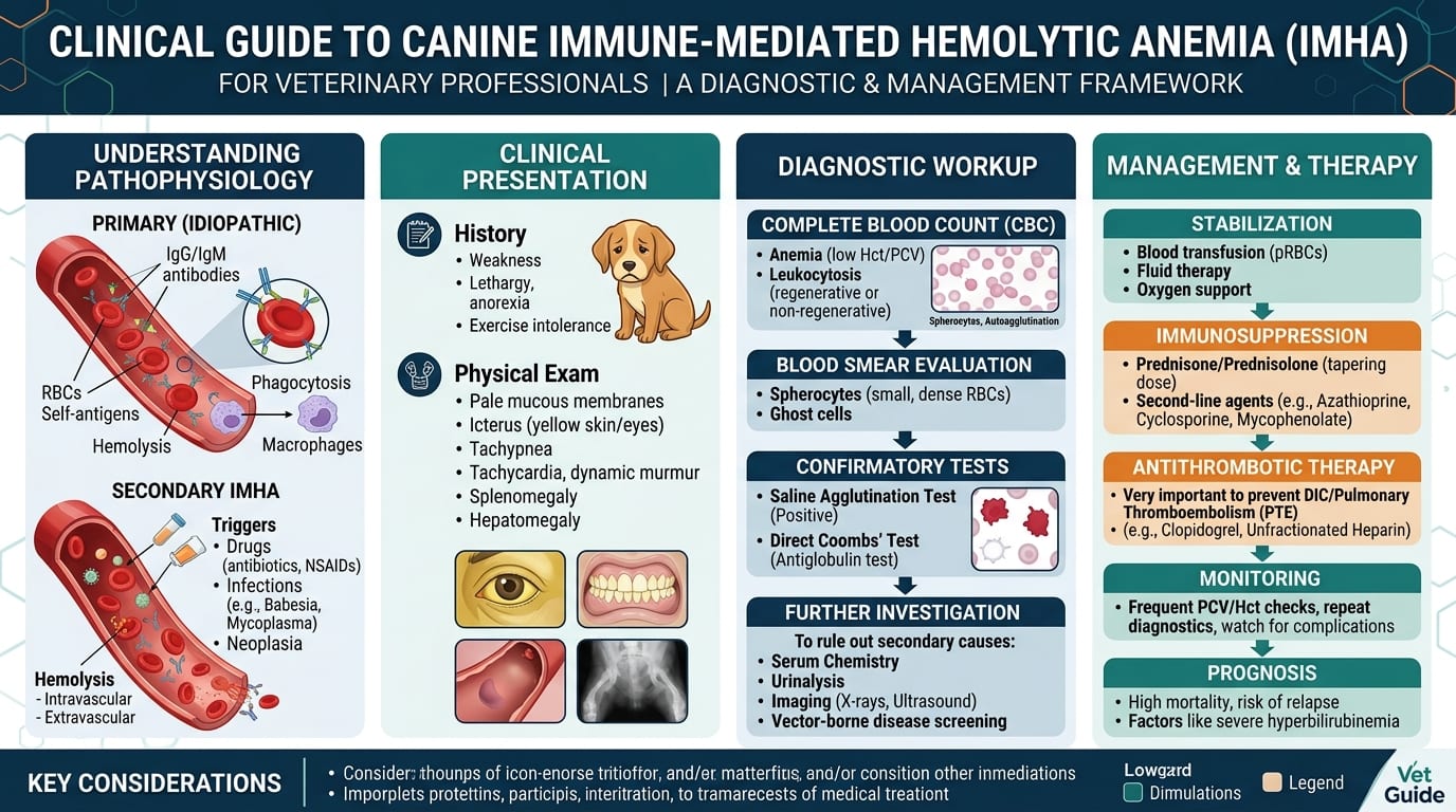
Immune-mediated hemolytic anemia (IMHA) in dogs is a life-threatening condition in which auto-antibodies destroy red blood cells. This guide covers the diagnostic workup (saline agglutination, Coombs test), PCV transfusion thresholds, immunosuppressive protocols with prednisolone and adjunctive agents, and critical anticoagulation to prevent pulmonary thromboembolism.
Key Takeaways
- ✓Primary IMHA accounts for 60-75% of cases; always test for tick-borne disease, neoplasia, and drug triggers as secondary causes.
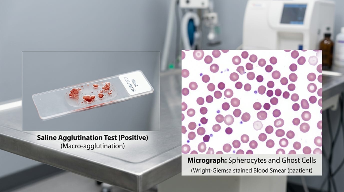
- ✓Saline slide agglutination (persistent macroscopic clumping after saline wash) is a rapid bedside diagnostic test for IMHA.
- ✓Prednisolone 1-2 mg/kg/day is the foundation of immunosuppression; add mycophenolate mofetil (10-20 mg/kg q12h) for severe or refractory cases.
- ✓Transfuse pRBCs at PCV < 12-15% with hemodynamic compromise; always crossmatch before transfusion.
- ✓Pulmonary thromboembolism kills 30-80% of fatal IMHA cases; start anticoagulation (heparin CRI or LMWH) plus ultralow-dose aspirin (0.5 mg/kg q24h).
- ✓Overall mortality is 20-40%; relapse occurs in 10-20% of survivors — complete immunosuppressive taper takes 3-6 months.
Diagnostic Workup: Saline Agglutination, Coombs Test and Workup for Underlying Cause
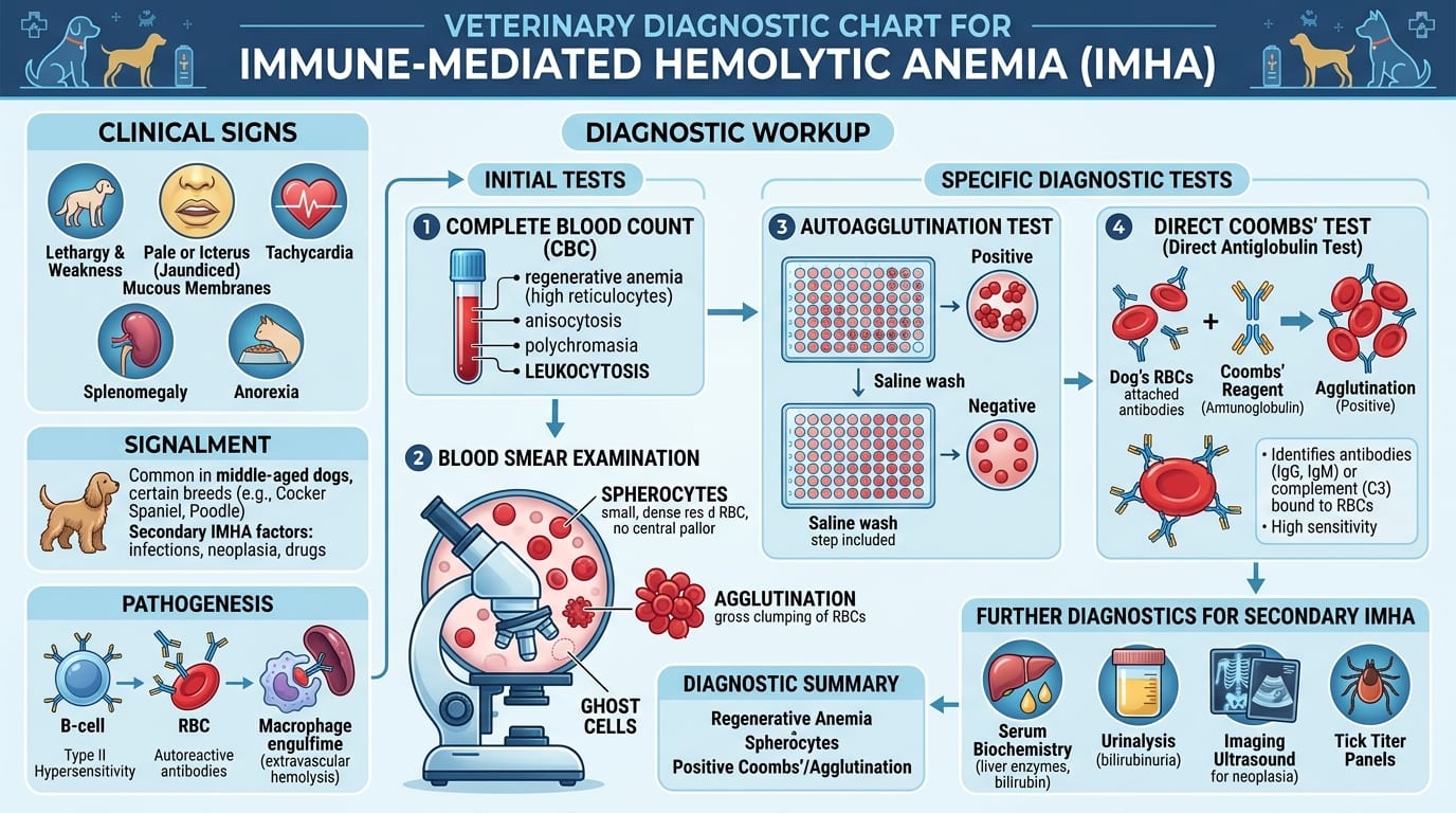
IMHA is classified as primary (idiopathic, autoimmune) in approximately 60-75% of cases, with the remainder secondary to infections, drugs, neoplasia, or vaccine reactions.
Predisposed Breeds
- American Cocker Spaniels, English Springer Spaniels
- Irish Setters, Poodles, Old English Sheepdogs
- Females more commonly affected than males
- Middle-aged dogs most common (peak 4-8 years)
Clinical Signs
- Pale (white/gray) or icteric mucous membranes
- Weakness, collapse, tachycardia, tachypnea (compensatory)
- Splenomegaly (extramedullary erythropoiesis)
- Bilirubinuria (dark urine), icterus
- Fever (hemolytic process + secondary infections)
Diagnostic Tests
*Saline Slide Agglutination (Bedside Test)*
- Mix 1 drop of EDTA blood with 1 drop of 0.9% NaCl on a glass slide
- True autoagglutination (macroscopic clumps) = confirms IMHA
- Rouleaux (coin stacking of RBCs) is a normal finding that washes away with saline; autoagglutination persists
*Complete Blood Count*
- PCV typically 10-25% (severe anemia); < 12% constitutes severe IMHA
- Regenerative anemia: polychromasia, reticulocytosis (> 1% reticulocytes), anisocytosis, macrocytosis
- Spherocytes (pathognomonic when numerous): small dense RBCs lacking central pallor
- Nucleated RBCs
- Thrombocytopenia in approximately 50-60% of IMHA cases (Evans syndrome if concurrent)
*Coombs Test (Direct Antiglobulin Test)*
- Detects IgG or IgM antibodies coating RBC surface
- Positive in approximately 60-90% of primary IMHA cases
- Negative Coombs does not rule out IMHA (false negatives if heavy hemolysis)
Workup for Underlying Cause
- Tick-borne disease PCR/serology (Ehrlichia, Anaplasma, Babesia)
- Blood culture (bacterial endocarditis, bacteremia-associated IMHA)
- Thoracic radiographs + abdominal ultrasound (neoplasia)
- Drug history (methimazole, cephalosporins, sulfonamides, NSAIDs can trigger)
- Vaccine-associated IMHA: most cases within 4 weeks of vaccination
Immunosuppressive Treatment Protocol and Blood Transfusion Thresholds
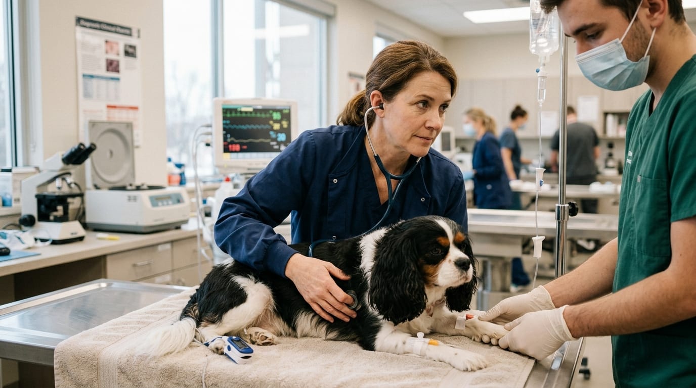
Immunosuppressive Therapy — First-Line
*Prednisolone or Prednisone*
- Dose: 1-2 mg/kg PO q24h (or divided q12h) as initial immunosuppression
- Begin at 2 mg/kg/day for severe or autoagglutinating IMHA
- Taper after PCV > 30% for 2-4 weeks: reduce by 25% every 4 weeks over 3-6 months
- Dexamethasone 0.3-0.5 mg/kg IV/IM q24h for initial IV immunosuppression in critical patients (equivalent anti-inflammatory dose without mineralocorticoid effects)
Adjunctive Immunosuppressants (Second-Line) Add when prednisolone alone insufficient or for steroid-sparing:
*Mycophenolate Mofetil (MMF)*
- Dose: 10-20 mg/kg PO q12h
- Preferred over azathioprine: faster onset (days vs. 4-6 weeks for azathioprine), fewer hepatotoxic effects
- Side effects: vomiting, diarrhea (give with food); rarely myelosuppression
- Monitor CBC every 2 weeks initially
*Azathioprine*
- Dose: 2 mg/kg PO q24h for 14-21 days, then every other day
- Hepatotoxic; baseline and monthly ALT/ALP monitoring required
- Onset of immunosuppression delayed 4-6 weeks
- TPMT enzyme deficiency in some breeds (Briard, Belgian Malinois) increases myelosuppression risk
Human IV Immunoglobulin (hIVIG)
- Dose: 0.5-1.0 g/kg IV over 6-12 hours
- Rapidly blocks Fc receptors on macrophages, reducing RBC destruction
- Expensive; reserved for severe autoagglutinating IMHA not responding to glucocorticoids
- Response in 24-72 hours
Blood Transfusion
*Transfusion Thresholds*
- PCV < 12-15% with clinical signs of hemodynamic compromise: transfusion indicated
- PCV 15-20% with severe autoagglutination and rapid decline: consider transfusion
- PCV > 20%: observe; transfusion generally not needed
- Blood typing (DEA 1.1) and crossmatch essential before transfusion
*Products*
- Packed red blood cells (pRBCs): 10-20 mL/kg IV over 4-6 hours; preferred over whole blood
- Fresh whole blood: useful if concurrent thrombocytopenia (Evans syndrome)
- Crossmatch essential, especially for previously transfused dogs (alloantibody risk)
*Post-Transfusion Monitoring*
- Measure PCV 1 hour post-transfusion and then every 12 hours
- Transfused cells will be destroyed by the same autoimmune process; maintain immunosuppression
Pulmonary Thromboembolism Prevention, Anticoagulation and Prognosis
Pulmonary Thromboembolism (PTE) — Leading Cause of Death in IMHA
- Activated platelets
- Procoagulant membrane phospholipids released from lysed RBCs
- Inflammatory state activating clotting cascade
- Glucocorticoid-induced hypercoagulability
Anticoagulation Protocols
*Heparin*
- Unfractionated heparin (UFH): 100-250 units/kg IV bolus, then 200-400 units/kg/hour CRI; target anti-Xa 0.35-0.70 IU/mL
- Alternatively: 150-250 units/kg SQ q6-8h (less predictable than CRI)
- Low molecular weight heparin (LMWH): dalteparin 100-150 IU/kg SQ q8-12h; enoxaparin 1.5 mg/kg SQ q12h — more predictable; anti-Xa monitoring optional
*Aspirin (Antiplatelet Therapy)*
- Ultralow-dose aspirin: 0.5 mg/kg PO q24h — provides antiplatelet effect without significant GI risk
- Often used in conjunction with heparin or as maintenance anticoagulation post-hospitalization
- Clopidogrel 1.1 mg/kg PO q24h can be used instead of aspirin
Monitoring Coagulation
- Prothrombin time (PT), activated partial thromboplastin time (aPTT)
- Thromboelastography (TEG) if available: hypercoagulable pattern in IMHA (shortened R time, increased angle, increased MA)
- D-dimers: elevated with ongoing fibrinolysis (hemolysis/thrombosis); > 0.5 mg/L warrants concern
Prognosis
- Overall mortality: 20-40% during first hospitalization
- Poor prognostic indicators: autoagglutination, bilirubin > 3.0 mg/dL, thrombocytopenia (Evans syndrome), neutrophilia > 30,000/uL, PTE
- Relapse: occurs in 10-20% of cases; may be triggered by infection, vaccination, stress
- Complete tapering of immunosuppression: typically 3-6 months; some dogs require maintenance low-dose prednisolone
Monitoring During Treatment
- PCV/TS every 12-24 hours during hospitalization
- Chemistry panel (ALT, bilirubin, BUN/creatinine) every 48-72 hours
- Peripheral blood smear review daily (spherocytes decreasing = good prognostic sign)
- Reticulocyte count every 72 hours (regeneration expected by day 3-5)
Frequently Asked Questions
What is the prednisolone dose for IMHA in dogs?
The initial immunosuppressive dose is 1-2 mg/kg PO q24h (or q12h for severe cases). Start at 2 mg/kg/day for severe or autoagglutinating IMHA. Once PCV is stable above 30% for 2-4 weeks, begin gradual tapering by 25% every 4 weeks over a total of 3-6 months.
When should a dog with IMHA receive a blood transfusion?
Transfusion is generally indicated when PCV drops below 12-15% with clinical signs of hemodynamic compromise (weakness, collapse, tachycardia). Dogs with PCV 15-20% and rapid RBC decline or severe autoagglutination should be evaluated individually. Packed RBCs (10-20 mL/kg over 4-6 hours) are preferred; always crossmatch before transfusion.
Why is pulmonary thromboembolism so dangerous in IMHA?
PTE is the leading cause of acute death in IMHA dogs, occurring in 30-80% of fatal cases. IMHA creates a hypercoagulable state from lysed RBC membranes, platelet activation, and glucocorticoid therapy. Anti-Xa monitoring during heparin infusion is recommended; ultralow-dose aspirin (0.5 mg/kg q24h) or low molecular weight heparin (dalteparin 100-150 IU/kg SQ q8-12h) helps reduce PTE risk.
Is mycophenolate or azathioprine better as a second immunosuppressant for IMHA?
Mycophenolate mofetil (10-20 mg/kg PO q12h) is preferred over azathioprine because it has a faster onset of immunosuppression (days versus 4-6 weeks), fewer hepatotoxic effects, and a more predictable side effect profile. Azathioprine (2 mg/kg PO q24h) requires regular ALT monitoring and is associated with bone marrow suppression in some breeds.
How is autoagglutination different from rouleaux on blood smear?
True autoagglutination (RBC clumping driven by IgM antibody bridging) persists when a drop of blood is diluted with 0.9% saline on a glass slide. Rouleaux (coin-stacking artifact from high protein or inflammation) disperses with saline. Macroscopic autoagglutination after saline wash is pathognomonic for IMHA.
References
- Swann JW, et al. ACVIM consensus statement on the diagnosis of immune-mediated hemolytic anemia in dogs and cats. J Vet Intern Med. 2019;33(2):313-334.
- Balch A, Mackin A. Canine immune-mediated hemolytic anemia: pathophysiology, clinical findings, and management. Compend Contin Educ Vet. 2007;29(4):217-225.
- Scott-Moncrieff JC, et al. Hemostatic abnormalities in dogs with primary immune-mediated hemolytic anemia. J Am Vet Med Assoc. 2001;218(12):1946-1951.