Quick Answer
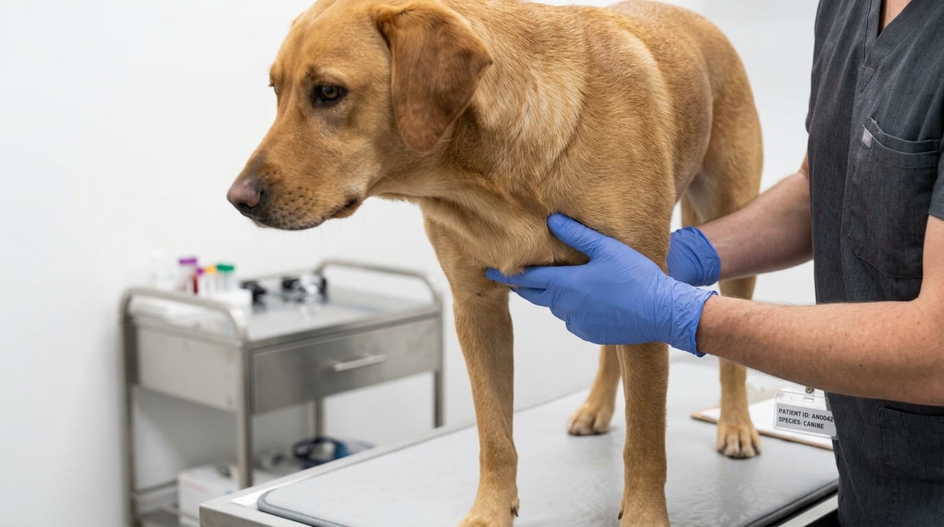
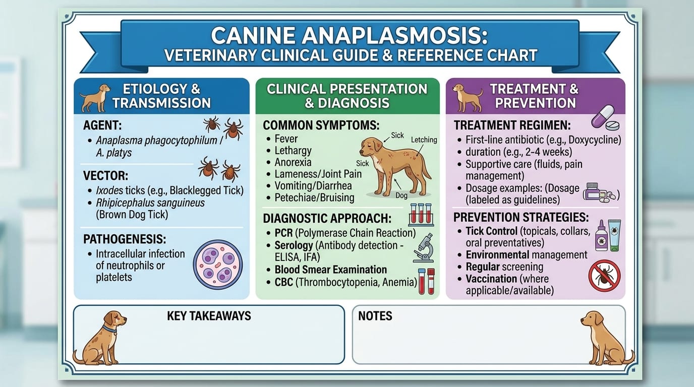
Anaplasmosis in dogs is caused by two species: Anaplasma phagocytophilum (granulocytic anaplasmosis, transmitted by Ixodes ticks) and Anaplasma platys (cyclic thrombocytopenia, transmitted by Rhipicephalus sanguineus). This guide covers clinical differentiation, CBC patterns, PCR diagnosis, and doxycycline treatment protocols.
Key Takeaways
- ✓A. phagocytophilum (Ixodes ticks) causes granulocytic anaplasmosis with acute fever, thrombocytopenia, and joint pain; A. platys (Rhipicephalus ticks) causes cyclic thrombocytopenia, often milder.
- ✓Morulae are visible in neutrophils (A. phagocytophilum) or platelets (A. platys) on blood smear in 20-30% of cases.
- ✓SNAP 4Dx Plus detects Anaplasma antibodies; PCR confirms active infection — run before initiating antibiotics for highest sensitivity.
- ✓Doxycycline 10 mg/kg PO q24h for 28 days is the treatment of choice; most dogs improve dramatically within 24-72 hours.
- ✓Co-infections are common: Ixodes ticks transmit A. phagocytophilum, Lyme, and Babesia simultaneously — test for all in endemic areas.
- ✓Year-round isoxazoline prevention (fluralaner, afoxolaner, sarolaner) kills Ixodes and Rhipicephalus ticks within 8-12 hours, limiting transmission.
A. phagocytophilum vs A. platys: Clinical and Epidemiologic Differences
Two distinct Anaplasma species cause disease in dogs, with different tick vectors, cell tropisms, geographic distributions, and clinical presentations.
Anaplasma phagocytophilum (Granulocytic Anaplasmosis)
- Vector: Ixodes scapularis (deer tick / black-legged tick) in northeastern/north-central US; Ixodes pacificus in western US
- Cell tropism: neutrophils and eosinophils
- Geographic distribution: endemic in northeastern US, upper Midwest, Pacific coast, northern Europe
- Clinical signs: acute fever (39.5-41.5 C), lethargy, anorexia, joint pain/reluctance to move, vomiting
- CBC: thrombocytopenia (50,000-150,000/uL), mild anemia, neutropenia or leukopenia
- Morulae: visible in neutrophils in approximately 20-30% of smears during acute phase
- Co-infection: highly significant — Ixodes ticks also transmit Borrelia burgdorferi (Lyme disease) and Babesia microti; 10-50% of A. phagocytophilum-positive dogs in endemic areas also seropositive for Lyme disease
Anaplasma platys (Canine Cyclic Thrombocytopenia)
- Vector: Rhipicephalus sanguineus (brown dog tick)
- Cell tropism: platelets (thrombocytes)
- Geographic distribution: warmer climates; southern US, Mediterranean, South America, Asia
- Clinical signs: often subclinical or mild; cyclic thrombocytopenia with 1-2 week intervals; petechiae, epistaxis, uveitis
- CBC: cyclic thrombocytopenia (nadirs as low as 5,000-20,000/uL), recovering to near normal between cycles
- Morulae: visible in platelets on blood smear; requires careful examination
- Less severe than A. phagocytophilum in most cases
| Feature | A. phagocytophilum | A. platys |
|---|---|---|
| Vector | Ixodes ticks | Rhipicephalus sanguineus |
| Cell type infected | Neutrophils | Platelets |
| Clinical severity | Moderate-severe acute | Usually mild, cyclic |
| Geographic | Northeastern/Midwest US | Warm climates |
| Co-infection risk | Lyme, Babesia | E. canis |
Diagnostic Testing: PCR, Serology and In-House 4Dx
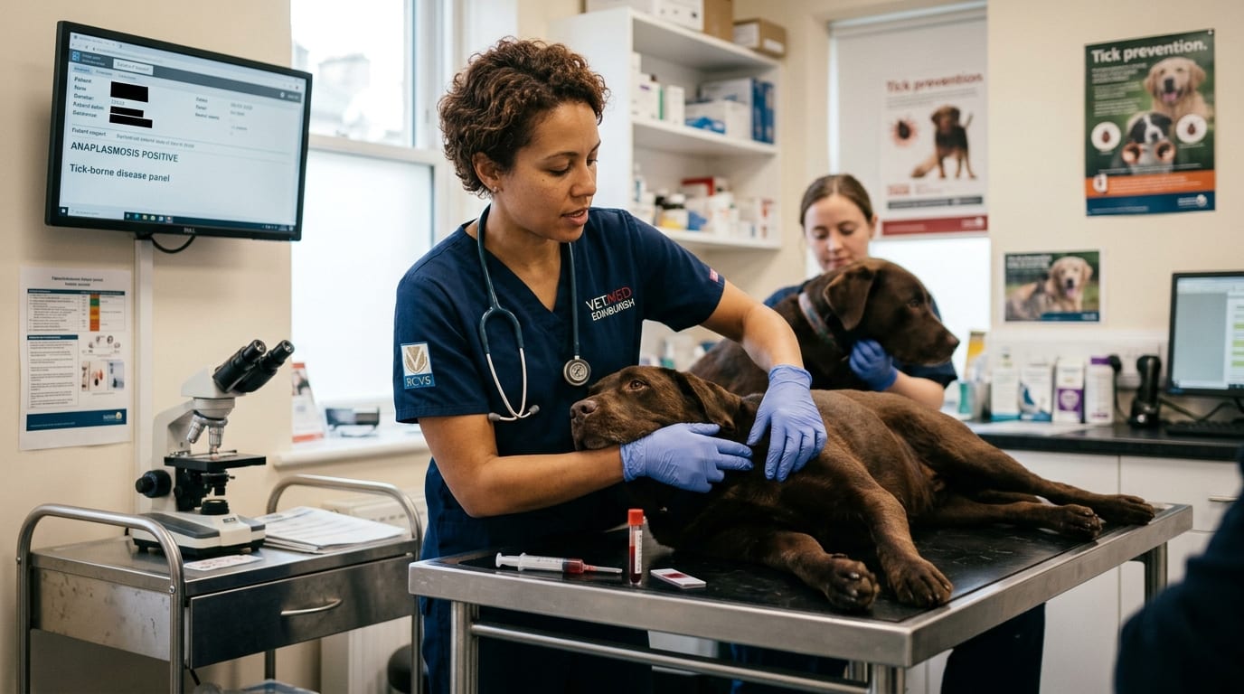
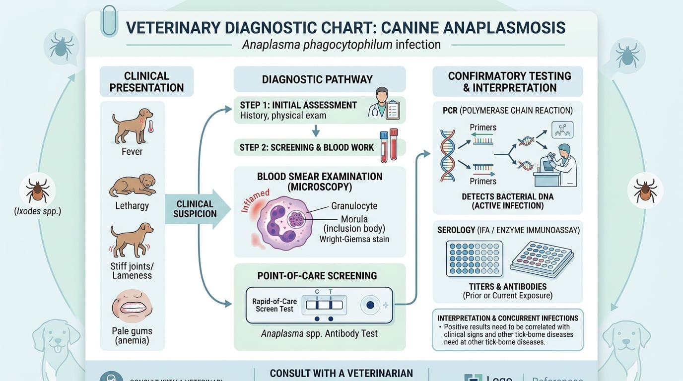
In-House SNAP 4Dx Plus (Antibody Detection)
- Antibodies develop 4-7 days after infection onset
- False negatives possible in acute infection before seroconversion
- Positive results in vaccinated-against-Lyme dogs may need differentiation
PCR (Definitive Diagnosis)
- PCR on EDTA blood is the most sensitive and specific test for active infection
- A. phagocytophilum PCR: highest sensitivity during acute phase (days 1-10)
- A. platys PCR: detects platelets-associated DNA; timing-dependent on platelet cycle
- Submit EDTA blood (1-2 mL) before antibiotic initiation for best results
Blood Smear Examination
- Examine 200-300 WBCs (for A. phagocytophilum in neutrophils)
- Examine platelet-rich areas (for A. platys in platelets)
- Sensitivity is low (20-30%); negative smear does not rule out infection
Treatment Protocol
- Doxycycline: 5-10 mg/kg PO q12h or 10 mg/kg PO q24h for 28 days
- Clinical improvement typically within 24-72 hours of initiating doxycycline
- Failure to respond promptly should prompt investigation for co-infections
Co-Infection Management
- A. phagocytophilum + Lyme disease: doxycycline treats both; add doxycycline 10 mg/kg/day
- A. phagocytophilum + Babesia: doxycycline does NOT treat Babesia; add imidocarb dipropionate 6.6 mg/kg IM once or twice (10-14 days apart)
- A. platys + E. canis: doxycycline treats both
Prognosis, Recurrence and Tick Prevention
Prognosis for A. phagocytophilum
- Excellent with prompt doxycycline treatment
- Most dogs recover fully within 1-2 weeks of treatment
- Severe thrombocytopenia with hemorrhage: transfusion may be needed; add blood products if PCV < 20% or platelets < 10,000/uL with active bleeding
- Immune-mediated sequelae (glomerulonephritis, immune-mediated thrombocytopenia) rare but possible
Prognosis for A. platys
- Most dogs recover completely with doxycycline
- Cyclic thrombocytopenia resolves within days of treatment initiation
- Recurrence uncommon with complete treatment course
Monitoring After Treatment
- CBC at day 14 and day 28 to confirm platelet recovery
- Serology may remain positive for months; do not use titer to assess cure
- If platelets do not normalize by day 14, reassess for co-infections or IMTP
Prevention
- Isoxazolines (fluralaner/Bravecto, afoxolaner/NexGard, sarolaner/Simparica): highly effective against both Ixodes and Rhipicephalus ticks; kill ticks within 8-12 hours
- Tick checks after outdoor exposure in endemic areas
- Vaccination against Lyme disease (Borrelia burgdorferi) for dogs at risk — does NOT protect against anaplasmosis
- Year-round prevention recommended in endemic areas (A. phagocytophilum in northeastern US; A. platys in southern US)
Zoonotic Potential
- A. phagocytophilum is zoonotic — causes human granulocytic anaplasmosis (HGA) in people bitten by the same Ixodes ticks
- Dogs are not directly infectious to humans but serve as sentinels for tick exposure
- Advise owners in endemic areas to use personal tick protection
Frequently Asked Questions
What is the difference between Anaplasma phagocytophilum and Anaplasma platys in dogs?
A. phagocytophilum causes granulocytic anaplasmosis by infecting neutrophils, is transmitted by Ixodes ticks, and causes acute fever, joint pain, and thrombocytopenia primarily in northeastern US. A. platys infects platelets, is transmitted by Rhipicephalus (brown dog tick), and causes cyclic thrombocytopenia with 1-2 week intervals, often in warmer climates.
Does SNAP 4Dx detect both Anaplasma species?
Yes. The IDEXX SNAP 4Dx Plus tests for antibodies to A. phagocytophilum antigen, but due to cross-reactive antigens, it also detects A. platys antibodies. A positive result indicates exposure; PCR is required for definitive active infection diagnosis.
How quickly do dogs improve with doxycycline for anaplasmosis?
Dogs with acute A. phagocytophilum anaplasmosis typically show dramatic clinical improvement within 24-72 hours of starting doxycycline 10 mg/kg q24h. Failure to improve within 48-72 hours should prompt testing for co-infections such as Babesia.
Is anaplasmosis in dogs contagious to humans?
Dogs infected with A. phagocytophilum are not directly infectious to humans. However, A. phagocytophilum is zoonotic — people can contract human granulocytic anaplasmosis from the same Ixodes ticks that infect dogs. Dogs serve as sentinels for tick exposure risk in endemic areas.
How long should doxycycline be given for anaplasmosis?
The standard treatment course is doxycycline 10 mg/kg PO q24h (or 5 mg/kg q12h) for 28 days. Some clinicians treat uncomplicated A. phagocytophilum for 14 days if rapid clinical response is achieved, though 28 days is recommended to ensure clearance.
References
- Carrade DD, et al. Canine granulocytic anaplasmosis: a review. J Vet Intern Med. 2009;23(6):1129-1141.
- Maggi RG, Breitschwerdt EB. Isolation of Candidatus Mycoplasma haematoparvum and Mycoplasma haemocanis from dog blood. Parasit Vectors. 2012;5:16.
- Kohn B, et al. Clinical features of natural Anaplasma phagocytophilum infection in dogs. J Vet Intern Med. 2008;22(6):1289-1295.