Quick Answer
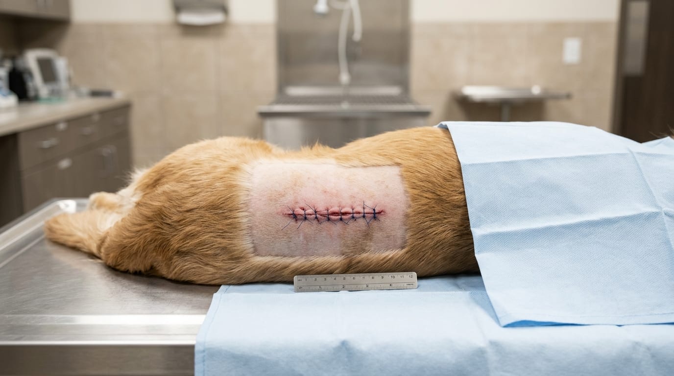
Effective wound management in dogs and cats requires systematic assessment, adequate lavage, appropriate debridement, and evidence-based closure timing. This guide covers wound contamination classification, lavage pressure and volume targets, debridement layers, and closure decision-making including primary, delayed primary, and second-intention healing.
Key Takeaways
- ✓Contaminated wounds older than 6 hours should be managed as open wounds initially; premature closure of contaminated wounds leads to abscess formation and dehiscence.
- ✓Lavage at 7–8 psi using a 35 mL syringe and 18-gauge needle with 50–100 mL isotonic saline per centimeter of wound length is the evidence-based decontamination standard.
- ✓Hydrogen peroxide, undiluted povidone-iodine, and alcohol are cytotoxic and must not be used for wound irrigation — they destroy granulation tissue and delay healing.
- ✓Dead space obliteration with buried absorbable sutures or active drain placement is essential to prevent seroma and abscess formation after closure.
- ✓Delayed primary closure at 3–5 days after initial open management is appropriate for contaminated wounds that have been adequately debrided and are forming clean granulation tissue.
- ✓Amoxicillin-clavulanate covers the primary wound pathogens in dogs and cats including Staphylococcus and Pasteurella; culture-directed therapy should be used for wounds failing to respond.
Wound Classification and Initial Assessment
Wound management begins with systematic classification that determines treatment urgency, closure timing, and antibiotic selection. The three-variable system — contamination level, wound age, and tissue viability — guides all subsequent decisions.
Contamination Classification
| Class | Definition | Closure |
|---|---|---|
| Clean | Surgical incision; no contamination | Primary |
| Clean-contaminated | Minor contamination; intact defense | Primary with irrigation |
| Contaminated | Fresh traumatic wound (<6h); gross soiling | Delayed primary or open |
| Dirty/infected | Old wound (>6h); purulent discharge; devitalized tissue | Open management; closure only after decontamination |
Wound Age and the Golden Period
- Clean traumatic wounds less than 4–6 hours old with minimal contamination can often be closed primarily after thorough irrigation
- Wounds older than 6 hours or with significant contamination should be managed as open wounds initially
Initial Assessment Checklist
- Estimate wound age and mechanism of injury
- Identify all wound margins (may be greater than visible — especially bite wounds and degloving injuries)
- Assess depth: skin, subcutaneous, muscle fascia, muscle belly, joint/body cavity involvement
- Check for foreign bodies: radiograph if penetrating object; ultrasound if soft tissue foreign body suspected
- Assess tissue viability: color, turgor, perfusion (capillary refill at wound margins, bleeding on pinch)
- Culture wounds older than 6 hours before antimicrobial therapy
Key Species Differences
- Cats: skin is very mobile; large dead space is common even with small wounds; Pasteurella multocida from cat bites requires specific antibiotic coverage
- Dogs: bite wounds may have extensive deep tissue damage with small surface punctures; explore all bite wounds surgically
Wound Lavage Pressure, Volume, and Debridement Techniques
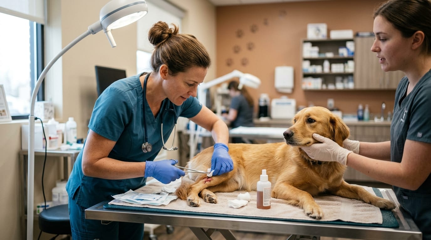
Lavage and debridement are the most important interventions for wound decontamination. Inadequate lavage is the most common reason for wound infection and dehiscence.
Lavage Technique
The goal of lavage is mechanical removal of bacteria, debris, and necrotic material without damaging viable tissue.
| Parameter | Recommendation |
|---|---|
| Pressure | 7–8 psi (pounds per square inch) — optimal for bacteria removal without tissue damage |
| Device | 35 mL syringe + 18-gauge needle = approximately 8 psi |
| Volume | 50–100 mL per cm of wound length (minimum) |
| Solution | 0.9% isotonic saline (preferred); tap water is acceptable if saline unavailable |
| Temperature | Warm saline (37°C) preferred to prevent hypothermia and maintain vasodilation |
Lavage Solutions: What NOT to Use
- Chlorhexidine >0.05% concentration: cytotoxic to fibroblasts; dilute 0.05% solution is acceptable
- Betadine (povidone-iodine) >0.1%: destroys fibroblasts at higher concentrations
- Hydrogen peroxide: cytotoxic; destroys granulation tissue; do not use
- Alcohol: cytotoxic; do not use
Debridement Methods
| Method | When to Use | Technique |
|---|---|---|
| Sharp debridement | Discrete necrotic tissue; tissue planes visible | Scalpel or scissors; excise layer-by-layer |
| En bloc debridement | Heavily contaminated wound with no viable tissue | Wide excision including all contaminated layers |
| Wet-to-dry dressings | Moderately contaminated; non-selective | Wet saline gauze applied and removed when dry — removes debris and non-viable tissue |
| Enzymatic (collagenase) | Selective; useful in delicate areas | Collagenase ointment applied topically |
| Surgical vacuum-assisted closure (VAC) | Large devitalized areas; delays primary closure | Negative pressure promotes granulation and removes exudate |
Tissue Viability Assessment During Debridement
- Pink, contractile muscle: viable — preserve
- Dark red/brown, non-contractile, non-bleeding muscle: devitalized — remove
- Pale, non-bleeding fat: devitalized — remove
- Hyperpigmented, crusty skin edges: eschar — debride to viable margins
Closure Timing: Primary, Delayed Primary, and Second-Intention Healing
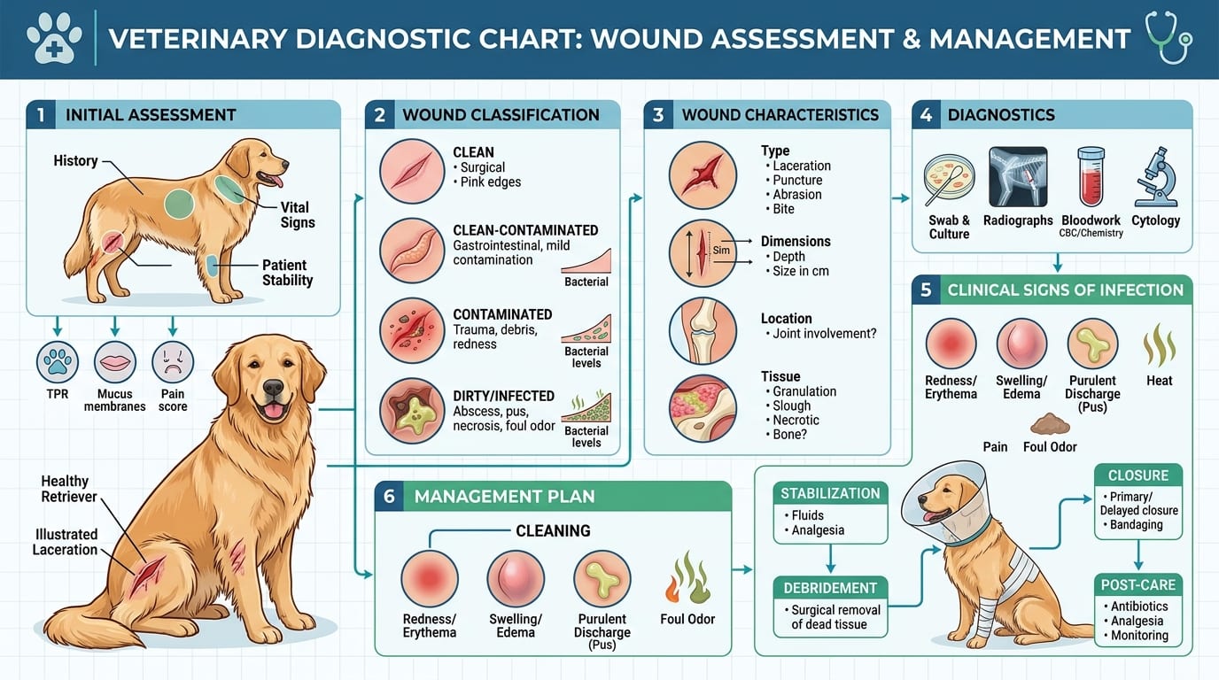
Closure timing is the single most consequential wound management decision. Premature closure of a contaminated wound results in abscess formation, dehiscence, and potentially systemic infection.
Closure Options
| Type | Timing | Indication |
|---|---|---|
| Primary closure | Immediately after wound preparation | Clean wounds; clean-contaminated wounds <6h old with adequate debridement |
| Delayed primary closure | 3–5 days after initial management | Contaminated wounds; wounds requiring initial open management |
| Secondary closure | After granulation tissue forms (5–14 days) | Wounds with tissue loss; second-intention healing underway |
| Second-intention | Wound heals open | Very large wounds unsuitable for closure; wounds with active infection |
When NOT to Close a Wound
- Active infection (purulent discharge)
- Tissue not viable after debridement
- Tension at wound margins (dog/cat skin does not stretch well)
- Dead space that cannot be obliterated
Suture Material Selection
| Layer | Suture | Pattern |
|---|---|---|
| Deep tissue/muscle | 3-0 or 2-0 PDS or Monocryl (absorbable monofilament) | Simple interrupted |
| Subcutaneous | 3-0 or 4-0 Monocryl | Buried interrupted to eliminate dead space |
| Skin | 3-0 Nylon or Prolene (non-absorbable) | Simple interrupted or cruciate |
Dead Space Management
- Obliterate with absorbable sutures in subcutaneous layers
- Penrose drain: passive drainage for large dead spaces; 3–5 days
- Jackson-Pratt drain: active suction for extensive dead space
Antimicrobial Selection for Wound Infections
| Organism | Drug of Choice |
|---|---|
| Staphylococcus spp. (dog bite) | Amoxicillin-clavulanate 12.5–25 mg/kg PO BID |
| Pasteurella multocida (cat bite) | Amoxicillin-clavulanate 12.5 mg/kg PO BID or doxycycline 10 mg/kg PO SID |
| Mixed contaminated wound | Amoxicillin-clavulanate + metronidazole 15 mg/kg PO BID |
| MRSA suspected | Culture-guided; doxycycline or chloramphenicol |
Frequently Asked Questions
What is the best solution for wound lavage in dogs and cats?
Isotonic 0.9% saline at 7–8 psi delivered with a 35 mL syringe and 18-gauge needle is the gold standard. Warm tap water is acceptable if saline is unavailable. Avoid hydrogen peroxide, undiluted betadine, or alcohol — all are cytotoxic to healing tissue.
How do you know if a wound can be sutured or needs to heal open?
Wounds older than 6 hours with significant contamination, devitalized tissue, or active infection should be managed as open wounds initially. After debridement and 3–5 days of open management, clean granulating wounds can be closed by delayed primary or secondary closure.
How do you treat a cat bite wound on a dog?
Cat bites create deep puncture wounds that introduce Pasteurella multocida into tissues. Explore all bite wounds surgically, lavage thoroughly, leave open or place a drain, and start amoxicillin-clavulanate immediately. Cat bites should never be underestimated — they frequently cause deep tissue infections.
What antibiotics should be used for infected wounds in dogs?
Amoxicillin-clavulanate (12.5–25 mg/kg PO BID) covers most wound pathogens including Staphylococcus and Pasteurella. For mixed contaminated wounds, add metronidazole (15 mg/kg BID) for anaerobic coverage. Culture-guided therapy is preferred for wounds not responding to first-line treatment.
How long should a drain remain in a wound?
Penrose drains are removed after 3–5 days once drainage volume decreases substantially. Jackson-Pratt drains are removed when daily output is less than 0.5 mL/kg/day. Leaving drains in place longer increases infection risk without additional benefit.
References
- Swaim SF, Henderson RA. Small Animal Wound Management. 3rd ed. Wiley-Blackwell; 2011.
- Bohling MW, Henderson RA, Swaim SF, et al. Cutaneous wound healing in the cat: a macroscopic description and comparison with cutaneous wound healing in the dog. Vet Surg. 2004;33(6):579-587.
- Robson MC, Heggers JP. Bacterial quantification of open wounds. Mil Med. 1969;134(1):19-24.