Quick Answer
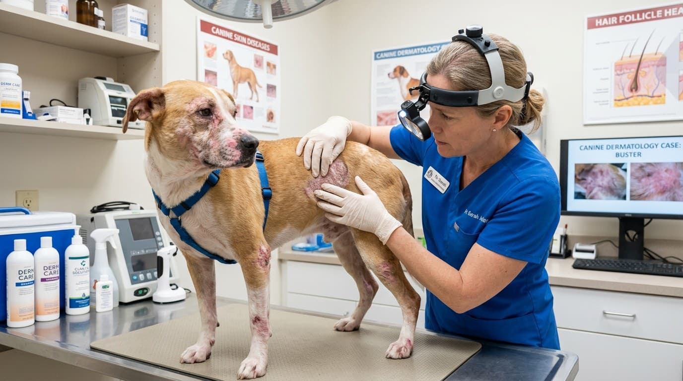
Mange is one of the most common and frequently misdiagnosed skin conditions in dogs, caused by two very different mites with different treatments and prognoses. This comprehensive guide covers sarcoptic mange (scabies), demodectic mange (red mange), diagnosis methods, and evidence-based treatment options.
Key Takeaways
- ✓Two distinct types of mange exist in dogs: sarcoptic (highly contagious, intensely pruritic, zoonotic) and demodectic (not contagious, immune-mediated, variable pruritus)
- ✓The pinnal-pedal reflex (rubbing ear edge triggers scratching) is a highly sensitive indicator of sarcoptic mange
- ✓Skin scraping sensitivity for sarcoptic mange is only 30–50%; empirical treatment trials are often more practical
- ✓Localized juvenile demodicosis has a 90% spontaneous remission rate; generalized demodicosis requires treatment and, in adults, investigation for underlying disease
- ✓Isoxazolines (NexGard, Simparica, Bravecto) are now first-line treatment for both types of mange — highly effective and well-tolerated
- ✓Never use corticosteroids in demodectic mange — they dramatically worsen mite burden
- ✓All contact dogs must be treated simultaneously for sarcoptic mange; environmental decontamination of bedding is also necessary
Understanding Mange: Two Very Different Diseases
The word "mange" describes any dermatological condition caused by parasitic mites. In dogs, two distinct types dominate clinical practice: sarcoptic mange and demodectic mange. Despite both being called "mange," they are caused by completely different mites, have different transmission routes, carry different prognoses, and require different treatments. Confusing the two — or using the wrong treatment — can have serious consequences.
Sarcoptic Mange (Canine Scabies)
- Caused by: *Sarcoptes scabiei var. canis*, an obligate burrowing mite
- Transmission: Highly contagious — spreads by direct contact with infected animals or contaminated fomites (bedding, grooming tools). Also temporarily zoonotic — humans exposed to infected dogs can develop a self-limiting, intensely itchy rash
- Immune component: Lesions are largely driven by hypersensitivity reaction to mite products, feces, and egg fragments — not just the mites themselves
- Key characteristic: INTENSE pruritus (itching), often described as disproportionate to the lesion severity visible
Demodectic Mange (Red Mange, Demodecosis)
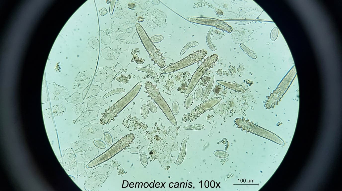
- Caused by: *Demodex canis* (and less commonly *D. injai*, *D. cornei*) — cigar-shaped mites that normally reside in small numbers in hair follicles
- Transmission: NOT directly contagious between dogs. Puppies acquire mites from their mother in the first days of life through close contact. Adult dogs with immune competence keep the mite population in check.
- Immune component: Disease occurs when the mite population proliferates unchecked — due to immunosuppression, genetic susceptibility (juvenile form), or underlying disease (adult-onset form)
- Key characteristic: Variable pruritus — may be minimal unless secondary bacterial infection occurs
Sarcoptic Mange: Signs, Diagnosis, and Treatment
Clinical Signs
Sarcoptic mange classically begins in areas of thin skin and sparse hair:
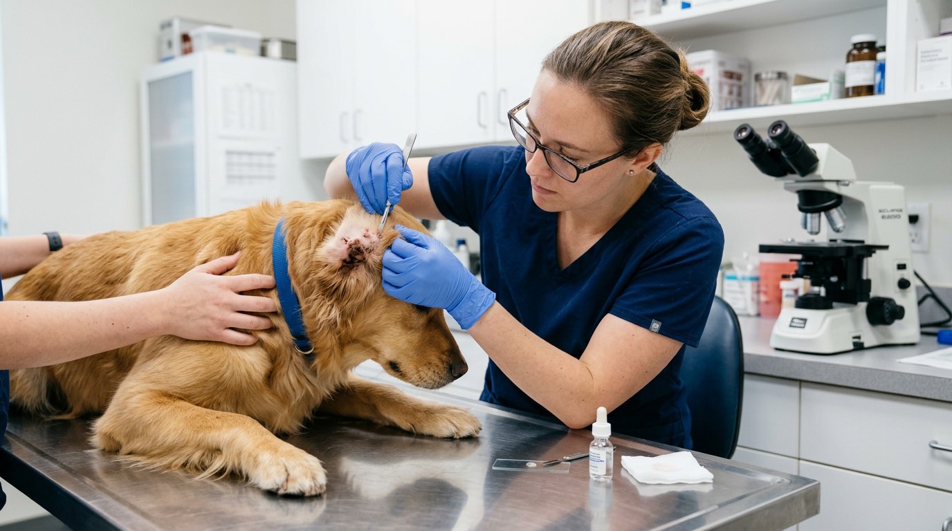
- Margins of the ear pinnae (ear flap edges) — the pinnal-pedal reflex is pathognomonic: when the ear margin is rubbed, the dog reflexively raises its hind leg to scratch — highly sensitive for scabies
- Elbows and hocks
- Ventral abdomen and chest
- Face and muzzle
As the disease progresses, the entire body can be affected. Intense pruritus is the hallmark — often more severe than the skin lesions suggest. Lesions include erythema, papules, crusts, scale, and in chronic cases, lichenification (skin thickening), hyperpigmentation, and secondary bacterial pyoderma or Malassezia overgrowth.
Zoonotic Importance
*S. scabiei var. canis* can temporarily infest humans in close contact with infected dogs, causing a papular, intensely itchy rash on the forearms, trunk, and skin contact areas. The mites cannot establish permanent breeding populations in humans (an "accidental host"), and the human rash typically self-resolves within 3–6 weeks once the dog is treated. However, the rash is severe enough to send owners to their own physicians — inform your doctor that your dog is being treated for scabies.
Diagnosis
Sarcoptic mange is notoriously difficult to confirm by skin scraping — the mites are present in low numbers and burrow deeply. Sensitivity of a single skin scraping is estimated at only 30–50%.
More reliable diagnostic approaches:
- Pinnal-pedal reflex: Positive in ~75–90% of dogs with scabies
- Serology (ELISA): Antibody testing against S. scabiei antigens — sensitivity ~80%, but takes 3–5 weeks post-exposure to become positive
- Empirical treatment trial: In dogs with classic history and clinical signs, treating for scabies and watching for improvement is often more diagnostically valuable than repeated negative scrapings
- Response to treatment remains the gold standard in practice
Treatment
Multiple highly effective options exist:
*Isoxazolines (first-line)*: Sarolaner (Simparica®), Afoxolaner (NexGard®), Fluralaner (Bravecto®), Lotilaner (Credelio®) — oral monthly to 12-week treatments that kill mites rapidly. All are highly effective against *S. scabiei*. Many dermatologists now use isoxazolines as first-line treatment due to ease of administration, palatability, and efficacy.
*Selamectin (Revolution®)*: Topical monthly application; effective but may require 2–3 applications 1 month apart.
*Doramectin / Ivermectin*: Injectable or oral; highly effective but off-label in dogs; AVOID in MDR1/ABCB1 mutation-positive breeds (Collies, Australian Shepherds, and related breeds) due to risk of neurological toxicity.
*Lime sulfur dips*: Older option; messy and malodorous but safe for very young puppies where other options are limited.
Environmental decontamination is important: wash all bedding in hot water, treat all contact dogs, and be aware that mites survive off-host for 36–48 hours in favorable conditions.
All dogs in the household should be treated simultaneously, even if asymptomatic.
Secondary pyoderma should be treated with appropriate antibiotics (culture-guided when possible).
Demodectic Mange: Localized vs. Generalized
Background: Why Demodex Becomes Disease
All dogs carry a small, normal population of *Demodex canis* mites in their hair follicles — this is a commensal relationship in immune-competent dogs. Demodicosis develops when the mite population proliferates beyond the immune system's ability to control it. Two very different clinical scenarios exist:
Localized Juvenile Demodicosis
- Age: Typically 3–18 months
- Presentation: 1–5 discrete patches of hair loss (alopecia), mild scale, and variable erythema — commonly on the face (periocular, perioral), forelegs, or paws
- Pruritus: Usually absent or mild (unless secondary infection develops)
- Prognosis: Excellent — approximately 90% of localized cases resolve spontaneously with supportive care as the immune system matures
- Treatment: Typically conservative monitoring; topical antiseptic shampoos (benzoyl peroxide, chlorhexidine); treat secondary infections. Aggressive treatment may not be needed.
Generalized Demodicosis
Defined as more than 5 lesions, or involvement of a whole body region, or involvement of two or more paws.
*Juvenile-onset generalized (under 18 months)*: Significant immunological deficit — likely genetic. Requires treatment. Associated with a roughly 50% spontaneous recovery rate with treatment.
*Adult-onset generalized*: ALWAYS investigate for an underlying cause — internal disease, endocrinopathy (hypothyroidism, hyperadrenocorticism), immunosuppressive medications (corticosteroids), or neoplasia. Treating demodex without addressing the underlying trigger leads to relapse.
Pododermatitis (Foot Involvement)
Demodex pododermatitis — infestation of the paw skin and interdigital spaces — is notoriously treatment-resistant, causes significant pain and lameness, and requires aggressive, prolonged therapy.
Clinical Signs of Generalized Demodex
- Multifocal to diffuse alopecia
- Comedones (blackheads) — follicular plugging by mite bodies
- Furunculosis (ruptured hair follicles causing deep bacterial infection) and cellulitis in severe cases
- Hyperpigmentation and lichenification in chronic cases
- Intense pruritus typically indicates secondary bacterial pyoderma or Malassezia
Diagnosis
- Deep skin scraping: More sensitive than in sarcoptic mange; *Demodex canis* is larger and more readily visualized. Positive = mites, eggs, larvae present in elevated numbers (normal: 1–2 mites per field; demodex = large numbers)
- Trichogram (hair pluck): Can identify mite infestation with less sample discomfort; good for paws or periocular areas difficult to scrape
- Skin biopsy: For deep pododermatitis where surface scraping may not reach; reveals follicular mites and granulomatous inflammation
Treatment
*Isoxazolines*: Afoxolaner (NexGard®), Sarolaner (Simparica®), Fluralaner (Bravecto®) are now considered first-line treatment for demodicosis by the ICADA (International Committee on Allergic Diseases of Animals) guidelines. Monthly or quarterly oral administration results in high cure rates (80–95%) with good tolerability. Treatment continues until two consecutive negative skin scrapings (typically 3–6 months).
*Amitraz dips* (Mitaban®): Historically first-line; now less commonly used due to need for professional application, potential for toxicity (sedation, hypotension), and availability of safer alternatives.
*Doramectin/Ivermectin*: Effective but MDR1-gene concern applies. ALWAYS test at-risk breeds before using.
Secondary bacterial pyoderma requires concurrent antibiotic therapy — culture and sensitivity testing is strongly recommended for deep infections.
Do NOT use corticosteroids in demodectic mange — they worsen immunosuppression and dramatically increase mite burden.
Monitoring Treatment and Preventing Relapse
Monitoring During Treatment
For generalized demodicosis, objective monitoring is essential:
- Perform skin scrapings every 4–6 weeks during treatment
- Count mites per field and track the ratio of live vs. dead mites
- Treatment success is defined as two consecutive monthly negative (or nearly negative) skin scrapings
- Continue treatment for at least one month AFTER the first negative scraping to reduce relapse risk
For sarcoptic mange, clinical improvement (reduced pruritus within 2–4 weeks) is the primary monitoring tool. Repeat serology is rarely needed. Persistent pruritus beyond 4–6 weeks of treatment warrants evaluation for reinfection or alternative diagnosis.
Common Reasons for Treatment Failure
- Incomplete household treatment (untreated contact dogs in sarcoptic mange)
- Owner non-compliance with dosing schedule
- Underlying immunosuppression not identified or addressed (adult-onset demodex)
- MDR1 gene mutation causing drug toxicity when ivermectin/doramectin used
- Concurrent allergic skin disease masking response
- Misdiagnosis (not mange at all)
Prevention
*Sarcoptic mange prevention*:
- Avoid contact with infected animals or wildlife (foxes are a significant sarcoptic mange reservoir)
- Monthly isoxazoline preventatives are highly effective; dogs on NexGard, Simparica, Bravecto, or Credelio for flea/tick prevention are also protected against scabies
- Quarantine and treat newly adopted dogs from unknown backgrounds before introducing to resident pets
*Demodectic mange prevention*:
- Avoid breeding from dogs with a history of generalized demodicosis — there is a strong heritable predisposition
- Avoid immunosuppressive medications (especially systemic steroids) without veterinary guidance in predisposed breeds
- Routine wellness care and prompt treatment of underlying diseases reduces adult-onset demodex risk
Frequently Asked Questions
Can I catch mange from my dog?
Only sarcoptic mange is zoonotic. *Sarcoptes scabiei var. canis* can temporarily infest humans causing an intensely itchy rash, but cannot establish permanent breeding populations in people. The human rash self-resolves within 3–6 weeks once the dog is treated. Demodectic mange is NOT transmissible to humans.
What is the fastest treatment for mange in dogs?
Isoxazoline medications (afoxolaner/NexGard, sarolaner/Simparica, fluralaner/Bravecto) are highly effective and fast-acting for both sarcoptic and demodectic mange. Most dogs with sarcoptic mange show significant pruritus reduction within 2–4 weeks. Demodectic mange requires 3–6 months of treatment to achieve two consecutive negative scrapings.
What does mange look like on a dog?
Sarcoptic mange: intense scratching, crusty red patches on ear edges, elbows, and abdomen; hair loss and skin thickening with chronicity. Demodectic mange: patchy or diffuse hair loss with minimal itching (unless secondary infection); blackheads (comedones) in hair follicles; commonly starts on the face and legs in young dogs.
Will my dog's hair grow back after mange?
Yes, in most cases. Once the mite infestation is controlled and secondary infection is treated, hair regrowth occurs over weeks to months. Severely affected skin with significant scarring may have incomplete regrowth. Young dogs with localized demodicosis typically regrow hair completely.
References
- Mueller RS, et al. "Treatment of Demodicosis in Dogs: 2011 Clinical Practice Guidelines." Veterinary Dermatology, 2012.
- ICADA. "Recommendations for the Management of Canine Demodicosis." Veterinary Dermatology, 2022.
- Beugnet F, et al. "Efficacy of Afoxolaner in Dogs with Generalized Demodicosis." Veterinary Parasitology, 2016.
- Moriello KA. "Zoonotic Skin Diseases of Dogs and Cats." Animal Health Research Reviews, 2003.
- Scott DW, Miller WH, Griffin CE. "Müller & Kirk's Small Animal Dermatology." 7th Edition. Elsevier, 2013.