Quick Answer
A detailed guide to canine ear infections covering otitis externa and media, common pathogens including Malassezia and Pseudomonas, ear cytology, medical and surgical treatment options, and prevention strategies.
Key Takeaways
- ✓Ear infections are almost always secondary to an underlying cause — allergic skin disease, hypothyroidism, or conformational factors — that must be addressed to prevent recurrence.
- ✓Ear cytology should be performed at every otitis visit to identify yeast vs bacteria vs mixed infection and guide targeted therapy.
- ✓Pseudomonas infections are the most challenging bacterial cause and require culture-guided antibiotic selection plus TrizEDTA flushes.
- ✓Chronic, end-stage otitis may require TECA-BO surgery, which is curative but results in hearing loss on the affected side.
- ✓Routine ear cleaning and allergy management are the cornerstones of preventing recurrent ear infections.
Understanding Canine Ear Infections
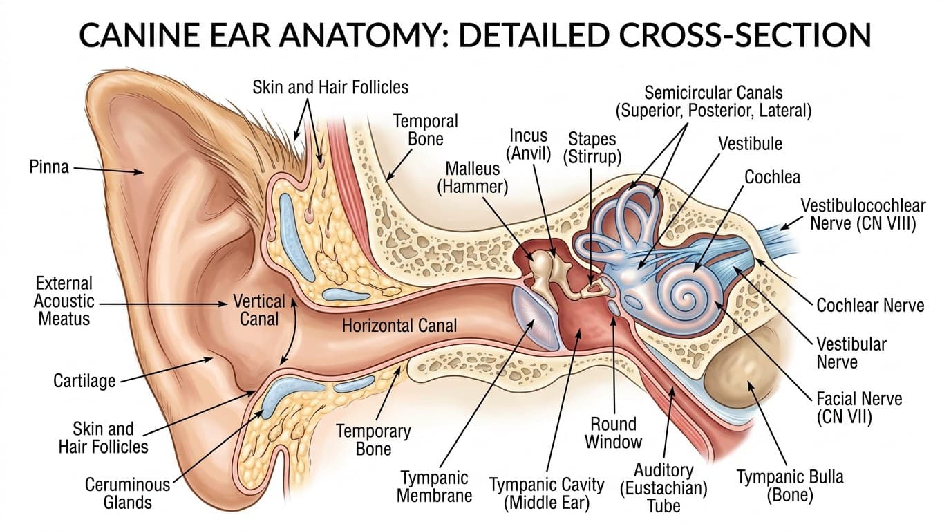
Otitis externa — inflammation of the external ear canal — is one of the most frequent reasons dog owners visit the veterinarian, affecting an estimated 10 to 20 percent of all dogs. The canine ear canal has a unique L-shaped anatomy (vertical canal descending, then turning horizontally toward the eardrum) that creates a warm, dark, humid environment ideally suited for microbial overgrowth when the normal ear ecosystem is disrupted.
Ear infections are classified by anatomical location. Otitis externa involves the external ear canal from the pinna to the tympanic membrane (eardrum). Otitis media involves the middle ear behind the eardrum and is present in an estimated 50 to 80 percent of chronic otitis externa cases, often undiagnosed. Otitis interna involves the inner ear and can cause vestibular signs (head tilt, circling, nystagmus).
It is crucial to understand that ear infections are almost always secondary to an underlying primary cause. Simply treating the infection without addressing the root cause guarantees recurrence. Primary causes include allergic skin disease (atopic dermatitis and food allergy — by far the most common), endocrine disorders (hypothyroidism, hyperadrenocorticism), conformational factors (narrow canals, excessive hair, pendulous ears), foreign bodies, and keratinization disorders.
Perpetuating factors — changes that develop as a consequence of chronic infection and prevent resolution — include progressive pathological changes to the canal such as stenosis (narrowing), calcification, and glandular hyperplasia. These structural changes can eventually make medical management impossible, necessitating surgical intervention.
Common Pathogens & Cytology
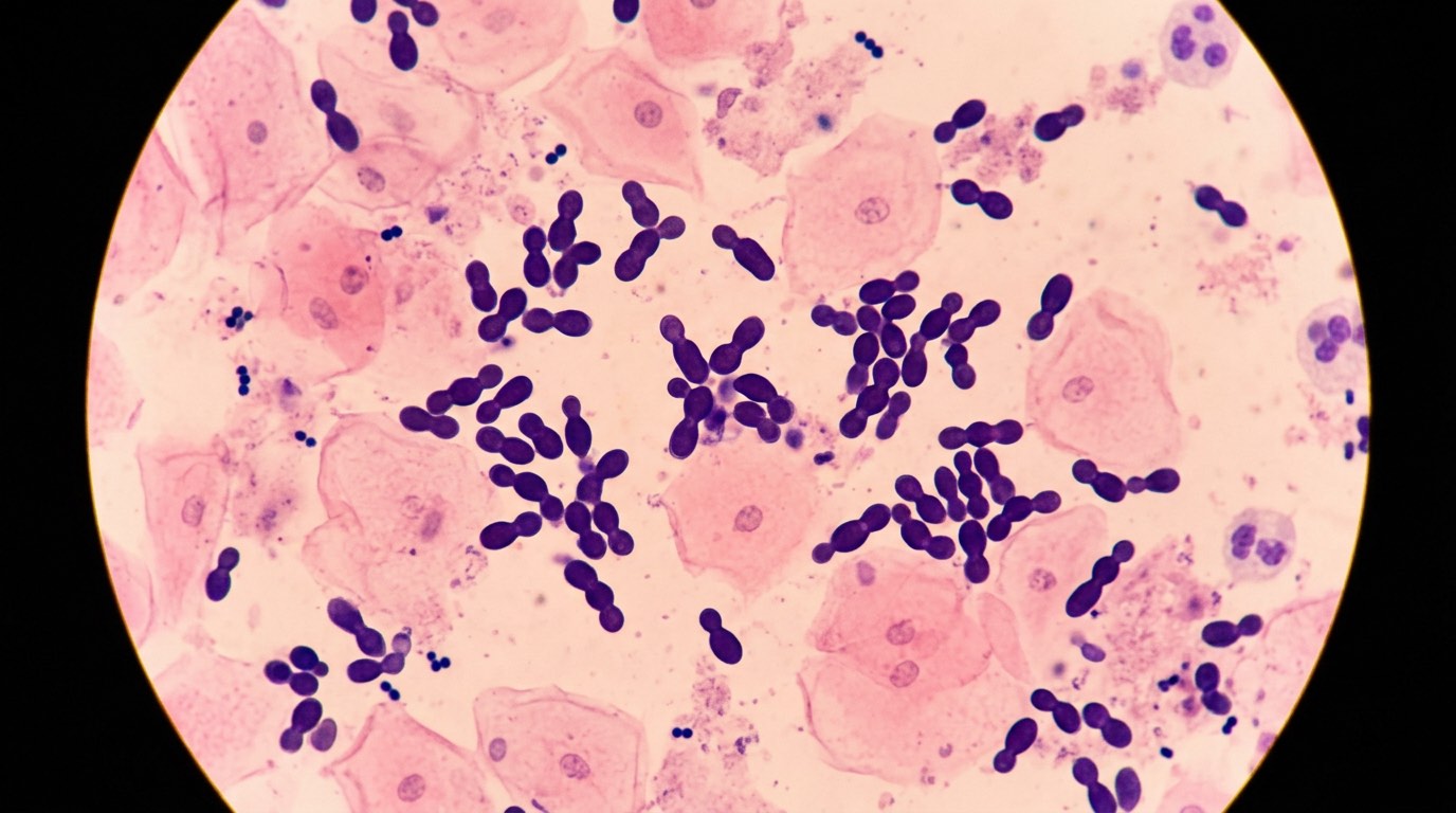
Ear cytology is a simple, inexpensive, and indispensable diagnostic tool that should be performed at every ear infection visit. A swab of ear debris is heat-fixed to a slide, stained, and examined microscopically to identify the types and quantities of organisms present, guiding targeted therapy.
Malassezia (Yeast)
Malassezia pachydermatis is the most common organism found in canine otitis externa. These peanut-shaped budding yeast organisms are easily recognized on cytology. Malassezia otitis typically produces a characteristic dark brown, waxy, sweet-smelling discharge. Yeast infections are frequently associated with underlying allergic disease and often respond well to topical antifungal therapy (miconazole, clotrimazole, ketoconazole).
Bacterial Infections
Cocci (round bacteria, usually Staphylococcus species) are the most common bacterial organisms in acute ear infections. They typically produce a yellow to tan purulent discharge and respond well to first-line topical antibiotics.
Rod-shaped bacteria — particularly Pseudomonas aeruginosa — represent a more challenging scenario. Pseudomonas infections produce a distinctive green-yellow, malodorous discharge and are notorious for antibiotic resistance. These infections frequently develop in chronically treated ears where repeated antibiotic courses have selected for resistant organisms. Culture and sensitivity testing is essential for Pseudomonas cases to guide effective antibiotic selection.
Mixed Infections
Many ear infections involve combinations of yeast and bacteria. Cytology guides the clinician in choosing combination products that address all organisms present, rather than treating blindly.
Ear Mites
Otodectes cynotis (ear mites) are more common in cats and puppies than adult dogs. They produce a characteristic dry, dark, coffee-ground-like debris. Mites can sometimes be visualized with otoscopy as tiny white moving dots. Modern isoxazoline flea/tick preventatives (NexGard, Simparica, Bravecto) are also effective against ear mites.
Medical Treatment
Successful treatment of ear infections requires three components: thorough ear cleaning, appropriate topical (and sometimes systemic) medication, and identification and management of the underlying primary cause.
Ear Cleaning
Professional ear cleaning — ideally under sedation for painful or severely infected ears — is the critical first step. Accumulated debris, pus, and biofilm must be removed before topical medications can contact the canal epithelium and be effective. Ceruminolytic agents (squalene-based cleaners like Cerumene) dissolve waxy debris, while aqueous-based cleaners (TrizEDTA) are preferred when the tympanic membrane may be compromised.
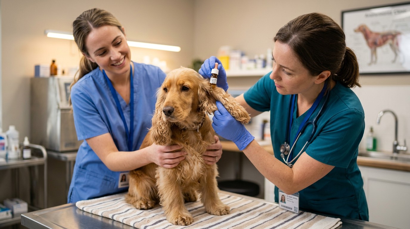
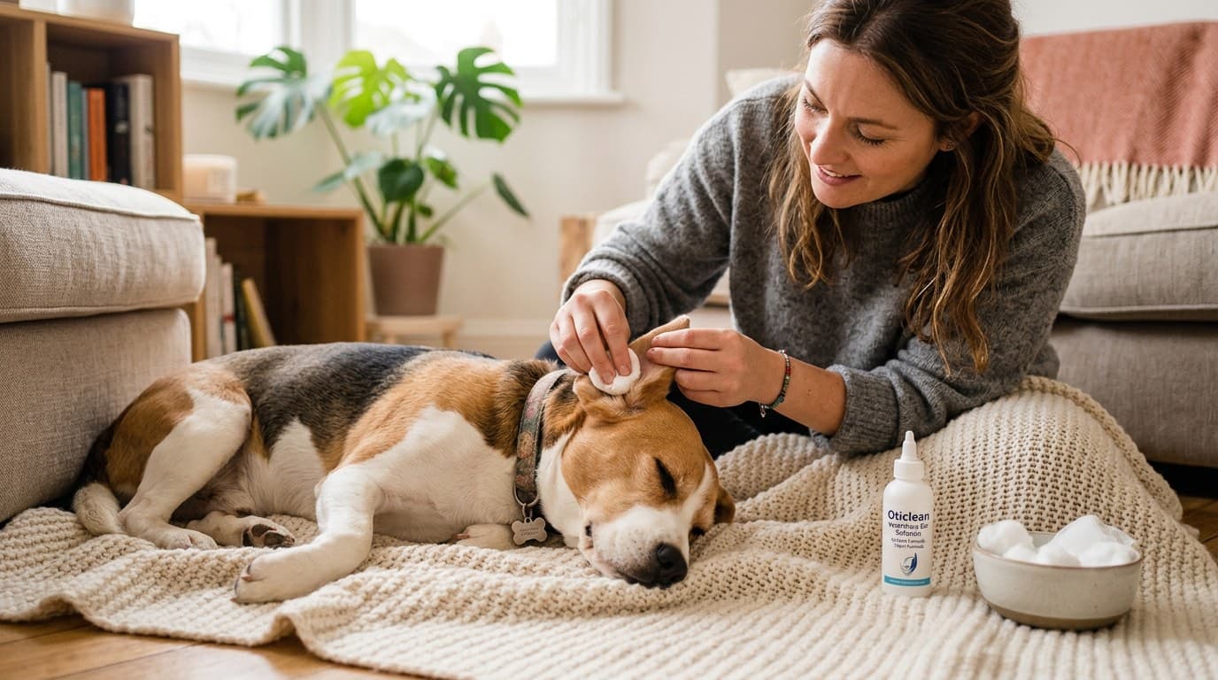
At home, owners should clean ears with a veterinarian-recommended solution before applying medication. The general technique involves filling the canal with cleaner, massaging the base of the ear for 30 seconds, allowing the dog to shake, and then wiping away loosened debris with cotton balls. Cotton swabs (Q-tips) should never be inserted into the ear canal.
Topical Medications
Most ear infections respond to topical therapy alone. Combination products typically contain:
- An antifungal (miconazole, clotrimazole) for yeast
- An antibiotic (gentamicin, marbofloxacin, florfenicol) for bacteria
- A corticosteroid (betamethasone, mometasone, dexamethasone) for inflammation and pain
For Pseudomonas infections, TrizEDTA flushes (which chelate divalent cations and disrupt bacterial cell membranes) applied 15–20 minutes before topical antibiotics significantly enhance antibiotic penetration and efficacy.
Systemic Therapy
Systemic antibiotics are reserved for otitis media (middle ear involvement), severe cases with periauricular cellulitis, and infections caused by highly resistant organisms. Fluoroquinolones (enrofloxacin, marbofloxacin) and extended courses based on culture results are often necessary.
Systemic antifungals (ketoconazole, itraconazole) may be needed for severe or refractory Malassezia infections, particularly those associated with generalized Malassezia dermatitis.
Chronic Otitis & Surgical Options
When ear infections recur repeatedly or fail to respond to aggressive medical management, the ear canal undergoes progressive pathological changes — epithelial hyperplasia, fibrosis, glandular hyperplasia, and eventually calcification and ossification of the cartilage. These changes narrow the canal, trap debris and moisture, and create a self-perpetuating cycle of infection that becomes increasingly refractory to medical therapy.
Lateral Ear Canal Resection (Zepp Procedure)
This procedure removes the lateral wall of the vertical ear canal to improve drainage and ventilation. It is most useful for cases where the primary problem is conformational (stenotic canals, excessive ear canal hair) without significant horizontal canal disease. Success rates are variable, and it does not address middle ear disease.
Total Ear Canal Ablation with Bulla Osteotomy (TECA-BO)
TECA-BO is the definitive surgical treatment for end-stage ear disease. The entire external ear canal is removed, and the tympanic bulla (middle ear cavity) is curetted clean. This procedure is curative for the ear infection but results in complete hearing loss on the operated side (which is typically already severely compromised in affected ears).
TECA-BO is a major surgery requiring general anesthesia, and potential complications include facial nerve damage (causing a drooped lip or inability to blink on the affected side), Horner's syndrome, para-aural abscessation, and wound dehiscence. Despite these risks, owner satisfaction is typically very high because the chronic pain, odor, and daily treatment burden are eliminated.
Video Otoscopy & Advanced Techniques
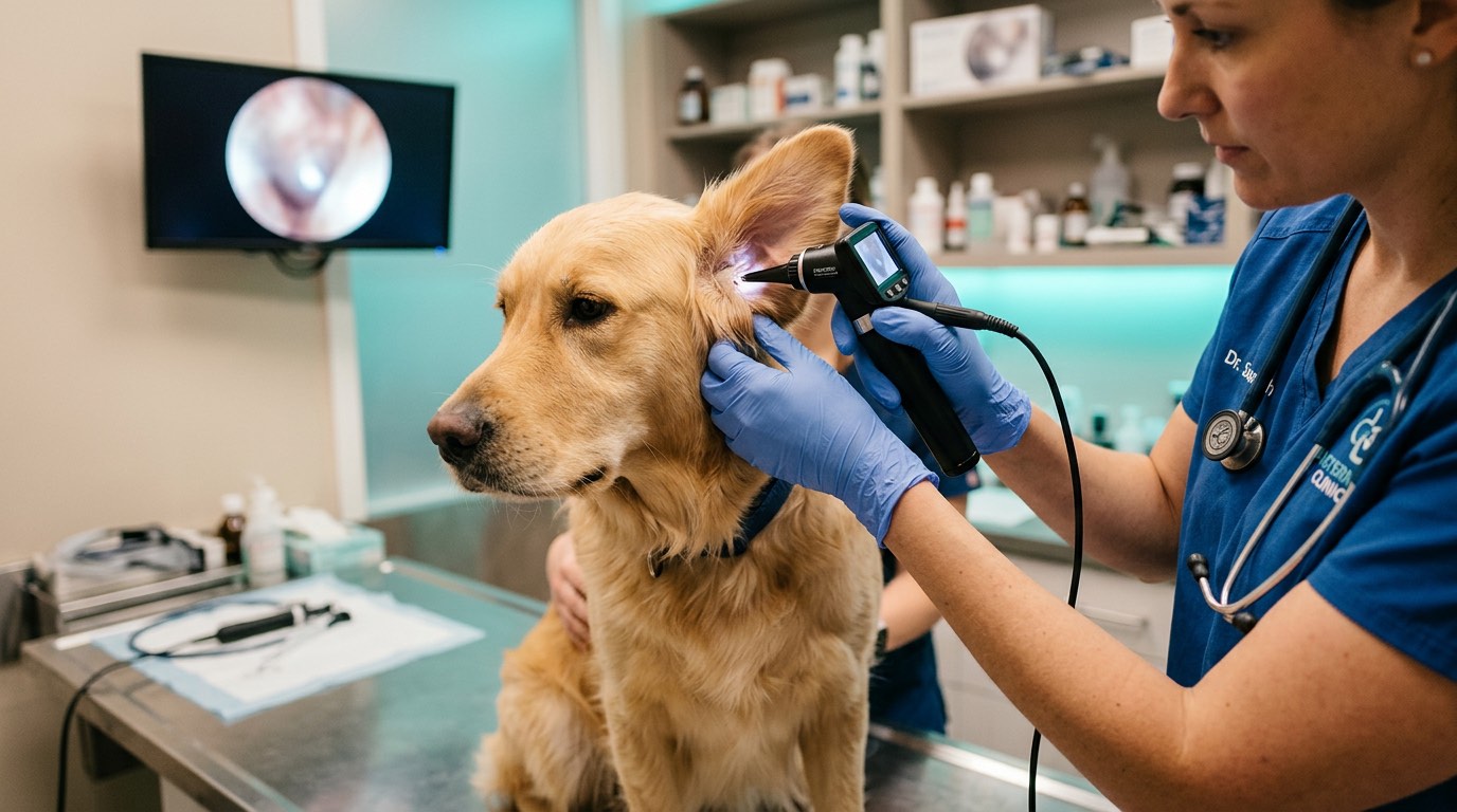
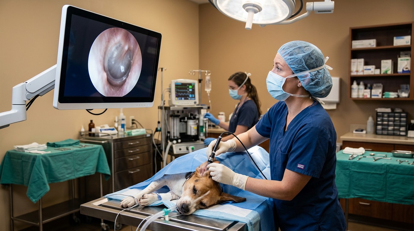
Video otoscopy allows magnified visualization deep into the ear canal and middle ear. It enables targeted flushing, foreign body removal, biopsy of masses, and myringotomy (deliberate puncture of the eardrum to sample and flush the middle ear). This technology has significantly improved the management of complex cases and often delays or avoids the need for surgery.
Prevention & Home Care
Preventing ear infections centers on controlling underlying primary causes and maintaining a healthy ear canal environment.
Managing the Underlying Cause
Since allergic skin disease is the most common primary cause of recurrent otitis, effective allergy management is the single most impactful preventive measure. This may include elimination diet trials to identify food allergies, allergen-specific immunotherapy (allergy shots) for environmental allergies, or daily medications such as oclacitinib (Apoquel) or lokivetmab (Cytopoint) to control allergic inflammation.
For dogs with hypothyroidism or other endocrine disorders, appropriate hormonal management reduces the frequency and severity of ear infections.
Routine Ear Maintenance
Dogs prone to ear infections benefit from regular ear cleaning — typically once weekly with a veterinarian-recommended ear cleaner. For swimming dogs, ears should be cleaned and dried after every water exposure. A drying ear cleaner containing a mild astringent helps maintain a less hospitable environment for yeast and bacteria.
Excessive ear hair in breeds like Poodles and Schnauzers was traditionally plucked, but current dermatology thinking suggests that routine plucking may actually cause more inflammation and should only be performed when hair is genuinely contributing to infection.
Warning Signs That Need Veterinary Attention
- Head shaking, ear scratching, or rubbing ears on furniture
- Odor, discharge, or redness visible in the ear
- Head tilt, circling, or loss of balance (suggesting middle or inner ear involvement)
- Swelling or pain when the ear base is touched
- Hearing loss or sudden behavioral changes
Early intervention — treating at the first sign of an ear problem rather than waiting for a full-blown infection — significantly improves outcomes and reduces the risk of chronic, irreversible ear canal changes.
Frequently Asked Questions
Why does my dog keep getting ear infections?
Recurrent ear infections almost always indicate an underlying primary cause, most commonly allergic skin disease (environmental or food allergies). Endocrine disorders like hypothyroidism can also predispose to chronic otitis. A thorough dermatological workup to identify and manage the root cause is essential for breaking the cycle.
Can I use hydrogen peroxide to clean my dog's ears?
Hydrogen peroxide is not recommended for ear cleaning because it can be irritating to inflamed tissue and may damage the delicate ear canal epithelium. Use a veterinarian-recommended ear cleaner specifically designed for dogs, which will have the appropriate pH and ingredients to safely dissolve debris without causing additional irritation.
How can I tell if my dog has an ear infection?
Common signs include head shaking, scratching at the ears, redness or swelling of the ear flap, odor coming from the ears, dark or colored discharge, and sensitivity or pain when the ear area is touched. Some dogs may tilt their head to one side if the infection is particularly painful or has progressed to the middle ear.
Are certain dog breeds more prone to ear infections?
Yes. Breeds with pendulous ears (Cocker Spaniels, Basset Hounds, Beagles), narrow ear canals (Shar Peis), excessive ear canal hair (Poodles, Schnauzers), or high rates of allergic disease (Bulldogs, Labrador Retrievers, West Highland White Terriers) are all predisposed to recurrent otitis.
What does it mean if my dog's ear infection won't go away despite treatment?
A non-resolving ear infection may indicate resistant organisms (especially Pseudomonas), undiagnosed middle ear involvement (otitis media), an unaddressed underlying primary cause like allergies, or progressive structural changes in the ear canal. A referral to a veterinary dermatologist for video otoscopy and advanced diagnostics is often warranted.
References
- Gotthelf LN. Diagnosis and treatment of otitis media in dogs and cats. Veterinary Clinics of North America: Small Animal Practice. 2004;34(2):469-487.
- Paterson S. Discovering the causes of otitis externa. In Practice. 2016;38(Suppl 2):7-11.
- Saridomichelakis MN, et al. Aetiology of canine otitis externa: a retrospective study of 100 cases. Veterinary Dermatology. 2007;18(5):341-347.