Quick Answer
A comprehensive guide to allergic conjunctivitis in dogs and cats, including causes, clinical signs, diagnosis, treatment options, and long-term management strategies.
Key Takeaways
- ✓Allergic conjunctivitis is inflammation of the eye membrane caused by immune overreaction to allergens
- ✓Common signs include redness, watery discharge, itching (pawing at eyes), and swelling
- ✓Diagnosis involves eye examination, cytology showing eosinophils, and possibly allergy testing
- ✓Always rule out corneal ulcers before using steroid eye drops (fluorescein test required)
- ✓Treatment includes antihistamine drops, mast cell stabilizers, cyclosporine, and allergen avoidance
- ✓Immunotherapy (allergy shots) may provide long-term improvement for environmental allergies
- ✓Prognosis is generally good with proper management, though lifelong treatment may be needed
What is Allergic Conjunctivitis?
Definition
Allergic conjunctivitis is inflammation of the conjunctiva (the thin, transparent membrane covering the white of the eye and inner eyelids) caused by an allergic reaction. It occurs when the immune system overreacts to allergens that come into contact with the eyes.
The Conjunctiva
The conjunctiva serves several important functions:
- Protects the eye from debris and microorganisms
- Produces components of the tear film
- Contains immune cells that respond to pathogens
- In allergic conditions, it becomes a site of excessive immune response
Types of Allergic Eye Disease
Type I Hypersensitivity (Immediate)
- Most common form
- Mediated by IgE antibodies and mast cells
- Occurs within minutes of allergen exposure
- Causes histamine release and inflammation
Type IV Hypersensitivity (Delayed)
- Cell-mediated immune response
- Takes hours to days to develop
- Often associated with contact allergens
Relationship to Other Allergic Conditions
Allergic conjunctivitis often occurs alongside:
- Atopic dermatitis
- Allergic rhinitis
- Food allergies
- Flea allergy dermatitis
- Seasonal allergies
In many cases, eye symptoms are part of a larger allergic syndrome affecting multiple body systems.
Causes and Risk Factors
Common Allergens
Environmental (Aeroallergens)
- Pollens (grass, tree, weed)
- Dust mites
- Mold spores
- Dander from other animals
Contact Allergens
- Household cleaning products
- Shampoos and grooming products
- Topical medications
- Plants (direct contact)
- Fabric or carpet fibers
Seasonal Patterns
- Spring/Summer: Tree and grass pollens
- Fall: Weed pollens (ragweed)
- Year-round: Dust mites, mold, indoor allergens
Some pets have perennial (year-round) symptoms with seasonal worsening.
Breed Predispositions
Dogs More Commonly Affected
- West Highland White Terriers
- Bulldogs (English, French)
- Labrador Retrievers
- Golden Retrievers
- Boxers
- Shar-Peis
- Dalmatians
- Most breeds with atopic dermatitis
Cats
- Less commonly diagnosed with primary allergic conjunctivitis
- Often occurs secondary to upper respiratory infections
- Siamese and Himalayan cats may be predisposed to atopy
Risk Factors
- Family history of atopic disease
- Living in highly pollinated areas
- Indoor allergen exposure
- Concurrent atopic dermatitis
- Age: Usually develops in young adults (1-3 years)
- Previous allergic conditions
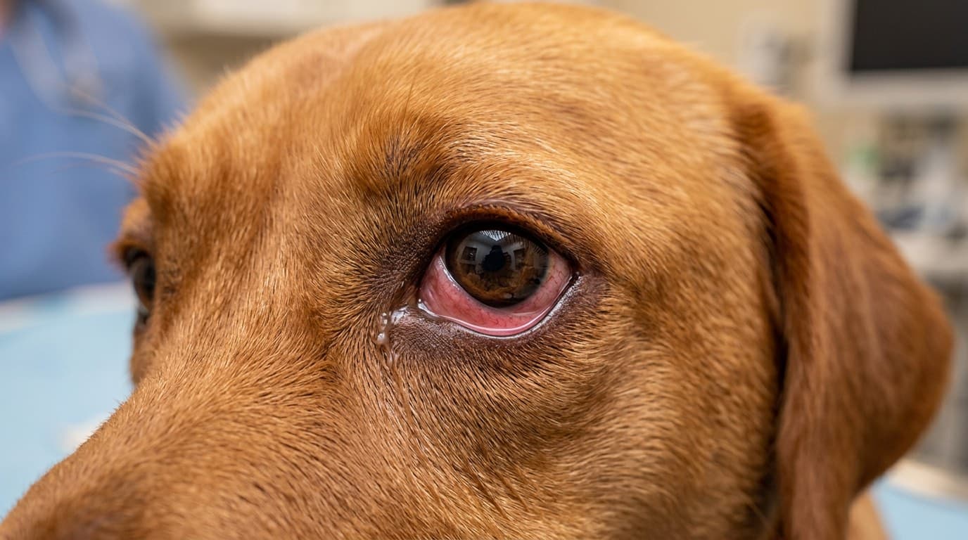
Clinical Signs
Common Symptoms
Eye Redness (Hyperemia)
- Pink to red discoloration of conjunctiva
- Visible blood vessels
- May affect one or both eyes
- Often worse with allergen exposure
Eye Discharge
- Clear, watery discharge (serous)
- May become mucoid (thicker, stringy)
- Usually not purulent (yellow/green) unless secondary infection
- Discharge may dry around eyes
Itching (Pruritis)
- Rubbing eyes with paws
- Rubbing face on carpet or furniture
- Squinting or blinking frequently
- Pawing at face
Swelling (Chemosis)
- Swollen, puffy conjunctiva
- May appear as fluid-filled bubble on eye surface
- Eyelids may be swollen
Tearing (Epiphora)
- Excessive tear production
- Tear staining beneath eyes
- Wet fur around eyes
Additional Signs
- Squinting (blepharospasm)
- Third eyelid elevation
- Photophobia (light sensitivity)
- Head shaking
- Concurrent skin symptoms (if atopic)
In Cats Specifically
- May develop conjunctivitis secondary to feline herpesvirus with allergic component
- Eosinophilic keratitis may occur
- Often have concurrent upper respiratory symptoms
Warning Signs Requiring Immediate Care
- Eye pain (squinting, avoiding light)
- Cloudy or blue-tinged cornea
- Change in pupil size
- Vision changes
- Yellow-green discharge (infection)
- Trauma to the eye

Diagnosis
Clinical History
Key questions to answer:
- Duration and pattern of symptoms
- Seasonal or year-round?
- Both eyes or one eye?
- Concurrent skin symptoms?
- Response to previous treatments?
- Environmental changes (new products, moving, etc.)?
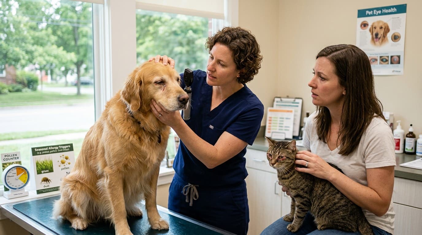
Ophthalmic Examination
Standard Eye Exam
- Visual inspection of eyes and adnexa
- Assessment of discharge character
- Evaluation of conjunctival appearance
- Corneal examination
- Pupil light responses
- Intraocular pressure if indicated
Schirmer Tear Test
- Measures tear production
- Rules out dry eye (keratoconjunctivitis sicca)
- Normal values vary by species
Fluorescein Staining
- Detects corneal ulcers or scratches
- Rules out corneal damage from rubbing
- Essential before prescribing steroids
Cytology
Conjunctival cytology can help identify:
- Eosinophils: Strongly suggests allergic cause
- Neutrophils: May indicate bacterial component
- Lymphocytes/Plasma cells: Chronic inflammation
- Bacteria: Secondary infection
- Mast cells may also be present
Allergy Testing
Intradermal Testing
- Gold standard for environmental allergies
- Requires specialist (dermatologist)
- Tests reaction to injected allergens
- Used to formulate immunotherapy
Serum Allergy Testing
- Blood test for allergen-specific IgE
- Less invasive than intradermal
- Can be performed by general practitioner
- Results used for immunotherapy
Elimination Diet Trial
- For suspected food-related allergies
- 8-12 week strict novel protein diet
- Helps identify food triggers
Ruling Out Other Causes
Must differentiate from:
- Bacterial conjunctivitis
- Viral conjunctivitis (especially in cats)
- Keratoconjunctivitis sicca (dry eye)
- Foreign body
- Eyelid abnormalities (entropion, ectropion)
- Corneal disease
- Uveitis
- Glaucoma
Treatment
Treatment Goals
- Relieve symptoms and discomfort
- Control inflammation
- Identify and avoid allergens when possible
- Prevent complications (corneal damage)
- Provide long-term management
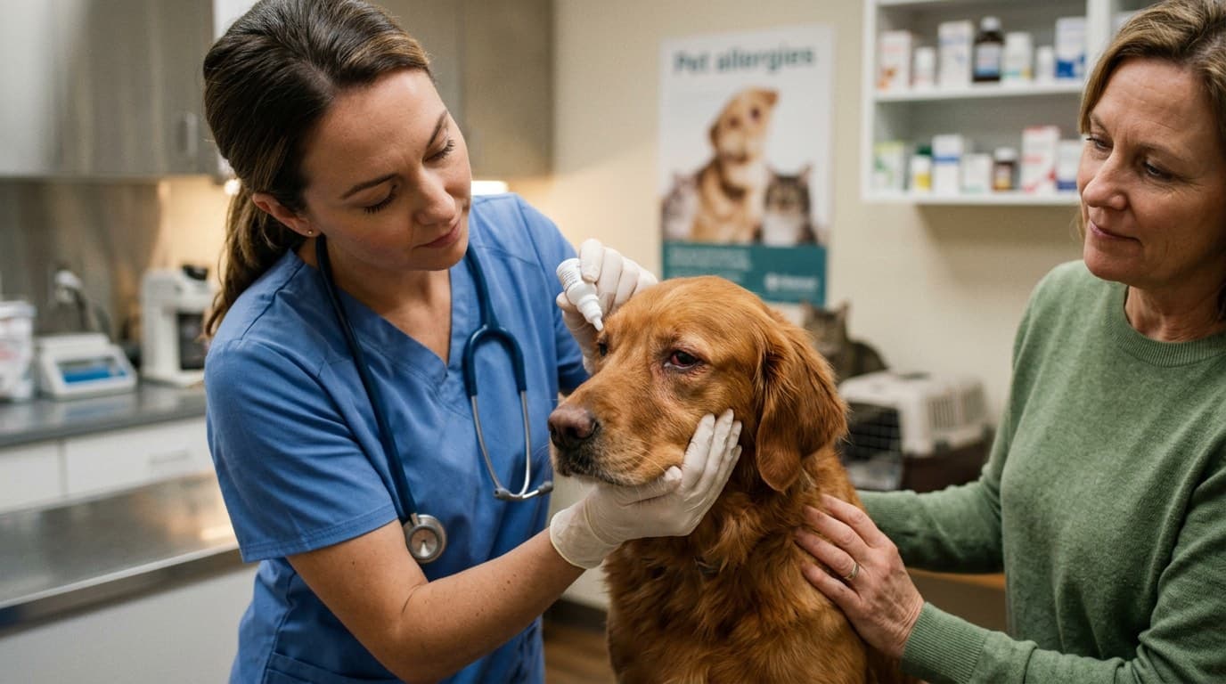
Topical Medications
Antihistamine Eye Drops
- First-line for mild cases
- Options: olopatadine, ketotifen
- Used 1-2 times daily
- Safe for long-term use
- May take 1-2 weeks for full effect
Mast Cell Stabilizers
- Prevent mast cell degranulation
- Options: lodoxamide, nedocromil
- Best used prophylactically before allergen exposure
- Can combine with antihistamines
Corticosteroid Eye Drops
- Very effective for inflammation
- Options: prednisolone acetate, dexamethasone
- NEVER use if corneal ulcer present (fluorescein test first!)
- Use short-term when possible
- May increase risk of infection
Cyclosporine/Tacrolimus
- Immunomodulatory agents
- Options: cyclosporine (Optimmune), tacrolimus
- Excellent for chronic management
- Takes 2-4 weeks for full effect
- Steroid-sparing properties
NSAID Eye Drops
- Anti-inflammatory without steroids
- Options: ketorolac, diclofenac
- Useful when steroids contraindicated
- May delay corneal healing
Artificial Tears
- Flush allergens from eye surface
- Provide lubrication
- Use preservative-free for frequent application
- Can apply 4-6 times daily as needed
Systemic Medications
Oral Antihistamines
- Help with concurrent allergic symptoms
- Options: diphenhydramine, cetirizine, hydroxyzine
- Variable efficacy for eye symptoms alone
- Safe for long-term use
Oral Corticosteroids
- Reserved for severe cases
- Short courses preferred
- Monitor for side effects
Apoquel (Oclacitinib) - Dogs
- Janus kinase inhibitor
- Very effective for atopic symptoms
- May help with ocular component
- Prescription only
Cytopoint (Lokivetmab) - Dogs
- Injectable anti-IL-31 antibody
- Monthly injection
- Targets itch pathway
- May provide some eye relief
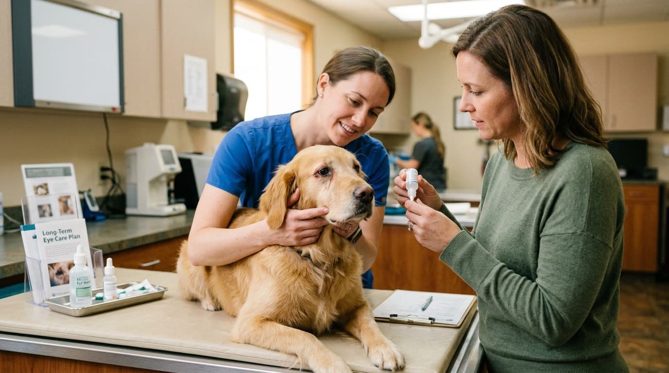
Long-Term Management
Allergen Avoidance
When allergens can be identified:
- Dust mites: HEPA filters, frequent cleaning, washable bedding
- Pollen: Limit outdoor time during peak seasons, wipe face/paws after outside
- Mold: Reduce humidity, clean high-moisture areas
- Contact allergens: Identify and remove specific products
Environmental Modifications
- Use air purifiers with HEPA filters
- Keep windows closed during high pollen times
- Wash pet bedding weekly in hot water
- Consider hypoallergenic beds
- Regular vacuuming with HEPA vacuum
- Limit carpeting in home
Bathing and Grooming
- Regular baths to remove allergens from coat
- Hypoallergenic, fragrance-free shampoos
- Wipe face and around eyes with damp cloth
- Consider omega fatty acid supplements for skin health
Immunotherapy (Allergy Shots/Drops)
Subcutaneous Immunotherapy (SCIT)
- Traditional allergy shots
- Gradual increase in allergen dose
- Modifies immune response over time
- Takes 6-12 months to see full effect
- 60-70% success rate
- Lifetime commitment in many cases
Sublingual Immunotherapy (SLIT)
- Allergen drops given under tongue daily
- Easier to administer than injections
- Similar efficacy to SCIT
- Growing option in veterinary medicine
Monitoring and Follow-Up
- Regular veterinary ophthalmic exams
- Monitor for secondary infections
- Adjust treatment based on seasonal patterns
- Watch for medication side effects
- Keep symptom diary
When to See an Ophthalmologist
- No improvement with standard treatment
- Recurrent corneal ulcers
- Severe or unusual presentation
- Suspected concurrent eye disease
- Chronic cases requiring advanced management
Complications and Prognosis
Potential Complications
Corneal Ulceration
- From chronic rubbing and scratching
- Can become infected or deep
- May require intensive treatment
- E-collar essential to prevent further trauma
Secondary Bacterial Infection
- Inflammation predisposes to infection
- Yellow-green discharge develops
- Requires antibiotic treatment
Keratoconjunctivitis Sicca (Dry Eye)
- Chronic inflammation can damage tear glands
- May develop secondary dry eye
- Requires lifelong tear supplementation
Chronic Corneal Changes
- Pigmentation of cornea
- Scarring
- May affect vision if central cornea involved
Eosinophilic Keratitis (Cats)
- White-pink plaque on cornea
- Associated with allergies and herpesvirus
- Requires specific treatment
Prognosis
For Allergic Conjunctivitis
- Generally good with appropriate management
- Most pets controlled with combination therapy
- Quality of life usually excellent
- May require lifelong treatment
- Symptoms often wax and wane with seasons
Factors Affecting Prognosis
- Ability to identify and avoid allergens
- Response to immunotherapy
- Compliance with treatment
- Presence of concurrent atopic dermatitis
- Development of complications
Quality of Life Considerations
Most pets with allergic conjunctivitis:
- Live normal, comfortable lives
- Enjoy normal activities
- Have minimal visual impact (usually)
- Require ongoing but manageable care
Owner Education
Key points for pet owners:
- This is usually a chronic, manageable condition
- Treatment may be lifelong
- Symptoms may flare seasonally
- Avoid rubbing (use E-collar if needed)
- Regular medication compliance is important
- Watch for signs of worsening or complications
Frequently Asked Questions
What causes allergic conjunctivitis in pets?
Common causes include environmental allergens (pollen, dust mites, mold), contact allergens (cleaning products, shampoos), and seasonal allergies. It often occurs alongside atopic dermatitis or other allergic conditions.
Can I use human eye drops on my dog or cat?
Never use human eye drops without veterinary guidance. Some human products contain ingredients toxic to pets. Always have a vet examine your pet first to rule out corneal ulcers, which can worsen with certain medications like steroids.
Is allergic conjunctivitis in pets curable?
While the underlying allergy cannot be cured, symptoms can be effectively managed with medications, allergen avoidance, and immunotherapy. Most pets maintain excellent quality of life with ongoing treatment.
How do I know if my pet's eye problem is allergies or an infection?
Allergic conjunctivitis typically causes clear/watery discharge, affects both eyes, and occurs seasonally or after allergen exposure. Infections often produce yellow-green discharge and may affect one eye. A veterinary exam with fluorescein staining and cytology can confirm the diagnosis.
References
- Maggs, D.J. et al. "Slatter's Fundamentals of Veterinary Ophthalmology." 6th Edition, Elsevier, 2018.
- Hendrix, D.V.H. "Diseases and Surgery of the Canine Conjunctiva and Nictitating Membrane." Veterinary Ophthalmology, 5th Edition, 2013.
- Miller, W.H. et al. "Muller and Kirk's Small Animal Dermatology." 7th Edition, Elsevier, 2013.
- Stiles, J. "Feline Herpesvirus." Clinical Techniques in Small Animal Practice, 2003.