Quick Answer
Feline HCM is the most common heart disease in cats. This guide covers breed-associated genetics (Maine Coon MYBPC3 mutation), echocardiographic diagnosis, medical management, and the recognition and emergency treatment of aortic thromboembolism (ATE).
Key Takeaways
- ✓HCM affects ~15% of cats; up to 33% of Maine Coons. MYBPC3 genetic testing available for Maine Coons and Ragdolls.
- ✓Echocardiographic diagnosis: LVFW or IVS thickness >6 mm diastole; LA:Ao >1.5 = high ATE risk
- ✓Gallop rhythm in a cat warrants immediate echocardiography — it is always pathological
- ✓CHF treatment: furosemide 2–4 mg/kg for acute edema; clopidogrel 18.75 mg/cat q24h for ATE prevention (superior to aspirin in FATCAT trial)
- ✓ATE emergency: acute hind-limb paralysis + cold limbs + absent femoral pulses — analgesia is the immediate priority
- ✓ATE prognosis: ~40–50% 30-day survival; return of motor function within 72 hours is the key prognostic indicator
What Is HCM? Pathophysiology and Prevalence
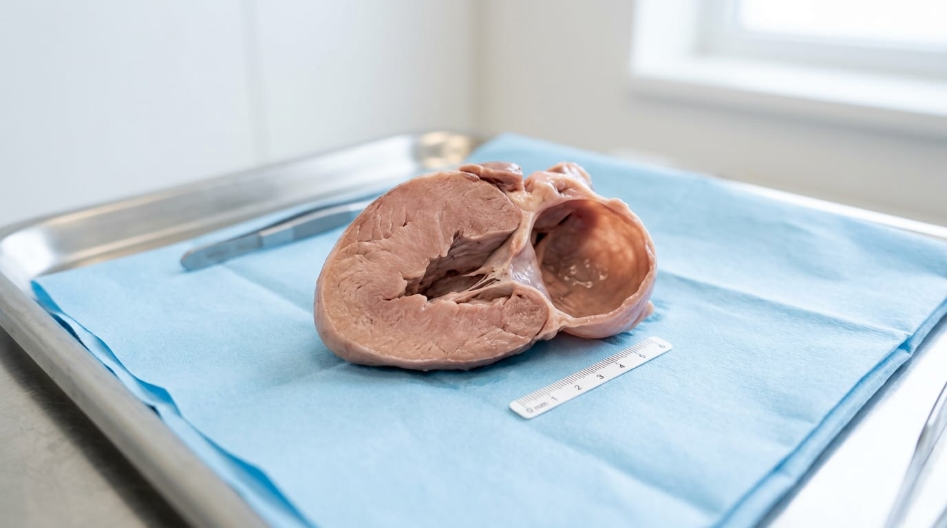
Hypertrophic cardiomyopathy (HCM) is characterized by abnormal hypertrophy of the left ventricular myocardium in the absence of other causes (hypertension, hyperthyroidism). It is the most common heart disease in cats.
Prevalence: Approximately 15% of the general cat population; up to 26–33% in Maine Coon and Ragdoll cats tested by echocardiography in research settings.
Pathophysiology The hypertrophied myocardium is stiff and poorly compliant (diastolic dysfunction). The left ventricle cannot relax and fill normally, leading to elevated filling pressures, left atrial enlargement, and eventually congestive heart failure (pulmonary edema, pleural effusion).
Secondary complications:
- Systolic Anterior Motion of the mitral valve (SAM): Hypertrophied papillary muscles and mitral valve leaflets obstruct the left ventricular outflow tract during systole — dynamic LVOTO. Creates a murmur and worsens hypertrophy.
- Left atrial enlargement: Dilated, poorly contractile LA → stasis of blood → thrombus formation → ATE risk
Genetic basis
- Maine Coon cats: Autosomal dominant mutations in MYBPC3 (cardiac myosin binding protein C). Two mutations identified: A31P and A74T. Genetic testing available.
- Ragdoll cats: A separate MYBPC3 mutation (R820W).
- Other breeds: Persians, British Shorthairs, Scottish Folds, Sphynx. Most cases in domestic shorthairs have unknown genetics.
Genetic test positive cats should not be bred and require regular echocardiographic monitoring.
Clinical Signs, Screening and Echocardiographic Diagnosis
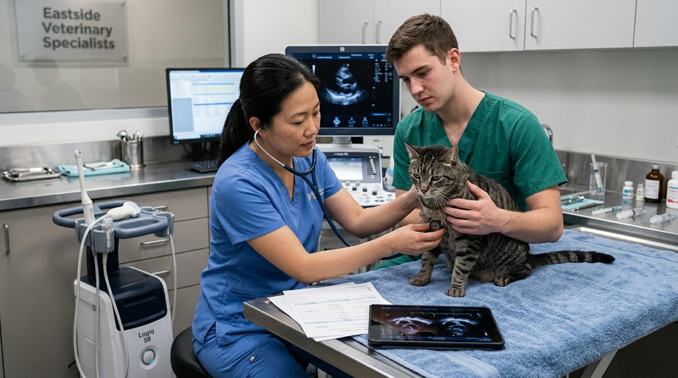
Clinical Signs Many cats with HCM are asymptomatic for years and are identified incidentally during routine examination.
Symptomatic signs:
- Dyspnea, open-mouth breathing, orthopnea — congestive heart failure (pulmonary edema)
- Pleural effusion: muffled heart/lung sounds ventrally, severely labored breathing
- Lethargy, anorexia, exercise intolerance
- Sudden hind-limb paralysis, cold limbs, vocalization — ATE (emergency)
Physical Examination
- Murmur (audible in ~50% of HCM cats due to SAM or turbulent flow) — Grade I–IV/VI
- Gallop rhythm (S3 or S4) — always pathological in cats; immediate echocardiogram recommended
- Tachycardia
- Muffled heart sounds (pleural effusion)
Screening Biomarkers
- NT-proBNP: Elevated in cats with significant cardiac disease. SNAP assay available (>100 pmol/L = significant cardiac disease). Useful for screening in dyspneic cats when echo is unavailable.
- Cardiac troponin I (cTnI): Elevated with myocardial injury. Prognostic marker.
Echocardiographic Diagnosis (Definitive)
- Diastolic left ventricular free wall (LVFW) or interventricular septal (IVS) thickness >6 mm = diagnostic for HCM (after excluding other causes)
- Left atrial to aortic root ratio (LA:Ao) >1.5 = significant left atrial enlargement (increased ATE risk)
- SAM of the mitral valve — dynamic LVOTO
- Doppler assessment of diastolic function (E/A ratio, tissue Doppler)
Echocardiography should be performed at referral level annually for cats at genetic risk.
Medical Management of HCM and CHF
Treatment goals: control signs of CHF, reduce ATE risk, minimize progression.
Preclinical HCM (no CHF signs)
- Atenolol (beta-blocker) 6.25–12.5 mg/cat PO BID: reduces heart rate, reduces dynamic LVOTO (SAM), may improve diastolic filling. Commonly used in cats with significant SAM or tachycardia.
- Diltiazem was historically used but is less favored over atenolol in current practice.
- No treatment has been proven to delay CHF onset in preclinical feline HCM (unlike in dogs with MMVD).
Acute CHF (Pulmonary Edema) Emergency management: 1. Oxygen supplementation (minimal handling — cats decompensate with stress) 2. Furosemide 2–4 mg/kg IV/IM/SQ (every 1–4 hours as needed until respiratory improvement) 3. Thoracocentesis if significant pleural effusion — diagnostic and therapeutic 4. Avoid fluids unless severely hypovolemic
Chronic CHF Management
- Furosemide 1–2 mg/kg PO BID–TID: primary diuretic. Monitor renal function and electrolytes.
- Atenolol 6.25 mg/cat PO BID: heart rate control, SAM reduction
- ACE inhibitors (enalapril, benazepril): modest benefit in some cats; commonly used despite limited evidence
- Torsemide: More potent loop diuretic; reserved for refractory cases
ATE Prevention (Antithrombotic Therapy)
- Clopidogrel (Plavix) 18.75 mg/cat PO q24h: Superior to aspirin for ATE prevention (FATCAT trial). First-line antithrombotic for cats with LA:Ao >1.5.
- Rivaroxaban (Xarelto) 2.5 mg/cat PO q24h: Direct Xa inhibitor; emerging evidence supports efficacy; may have lower re-ATE risk than clopidogrel in some studies.
Aortic Thromboembolism (ATE): Saddle Thrombus Emergency
ATE is one of the most devastating and acute emergencies in feline medicine. Thrombi form in the enlarged left atrium and embolize to the aortic trifurcation (saddle thrombus), blocking blood flow to the hind limbs.
Clinical Signs of ATE — Recognize Immediately
- Acute hind-limb paralysis or paresis (sudden onset, often minutes)
- Pain — extreme; cats vocalize, pant, or cry
- Cold, cyanotic hind limbs — firm, cool to the touch; femoral pulses absent or weak
- Nail bed cyanosis (pink → white/blue in affected limbs)
- Possible forelimb involvement if thrombus is more cranial
- Dyspnea often concurrent (due to underlying CHF)
Diagnosis Clinical presentation is usually diagnostic. Doppler probe: absent or significantly reduced arterial flow in femoral artery.
Emergency Management 1. Analgesia (PRIORITY): Buprenorphine 0.01–0.02 mg/kg buccal q6–8h; butorphanol IM; or low-dose fentanyl CRI 2. Manage concurrent CHF (oxygen, furosemide) 3. Heparin anticoagulation: unfractionated heparin 200–300 U/kg IV/SQ loading dose, then 150–300 U/kg SQ TID (helps prevent further thrombosis, not thrombolysis) 4. Warming of affected limbs (external warmers, blankets — avoid excessive heat) 5. DO NOT administer thrombolytics (streptokinase, tPA) routinely — high risk of fatal reperfusion injury 6. Supportive: IV fluids judiciously (avoid fluid overload in CHF cats), nutritional support
Prognosis
- 30-day survival with treatment: ~40–50%
- Cats with return of motor function within 72 hours have the best prognosis
- 6-month survival of discharged cats: ~60–70%
- Recurrence without antithrombotic therapy: >50% within 1 year
- Immediately initiate clopidogrel at discharge
References
- Payne JR, et al. Prognostic indicators in cats with hypertrophic cardiomyopathy. J Vet Intern Med. 2010.
- Hogan DF, et al. Secondary prevention of cardiogenic arterial thromboembolism in the cat (FATCAT study). J Vet Cardiol. 2015.
- Borgeat K, et al. Arterial thromboembolism in 250 cats in general practice. J Vet Intern Med. 2014.