Quick Answer
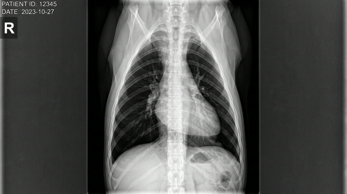
Pneumothorax is free air in the pleural space causing lung collapse and respiratory failure. This guide covers traumatic vs spontaneous causes, tension pneumothorax as an immediate emergency, needle decompression and chest drain placement, and surgical bullae resection.
Key Takeaways
- ✓Tension pneumothorax: immediate needle decompression without waiting for radiographs
- ✓Absent dorsal sounds and hyperresonance distinguish pneumothorax from pleural effusion (absent ventral)
- ✓Spontaneous PTX from bullae recurs >50% without surgery; refer for thoracoscopic resection
- ✓Chest tube with Heimlich valve is standard treatment for significant pneumothorax
- ✓Traumatic PTX: always check for concurrent contusions, hemothorax, diaphragmatic hernia
Types and Emergency Recognition
Classification
Traumatic Pneumothorax
- Most common type; MVA, bite wounds, blunt trauma
- Often bilateral (50-60% of trauma cases)
- Usually self-limiting if no ongoing air leak
Spontaneous Pneumothorax
- Secondary (most common): pulmonary bullae/blebs (80%), lung abscess rupture, neoplasia, parasitic cysts
- Primary (rare): no underlying disease
Tension Pneumothorax -- LIFE-THREATENING EMERGENCY One-way valve effect: air enters but cannot escape. Progressive pressure compresses heart and great vessels.
*Recognition:*
- Severe rapid respiratory deterioration
- Absent breath sounds bilaterally; cardiovascular collapse
- Tracheal deviation on radiograph
IMMEDIATE ACTION: 1. Do NOT wait for radiographs 2. Insert 14-16 gauge catheter at 2nd-3rd ICS dorsal thorax 3. Air rush confirms and treats simultaneously 4. Immediately follow with chest tube
Physical Exam Distinction
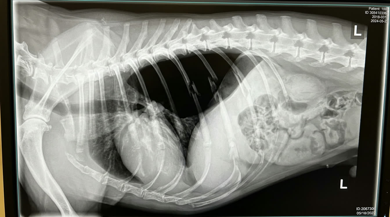
- Pneumothorax: absent breath sounds DORSALLY (air rises)
- Pleural effusion: absent breath sounds VENTRALLY (fluid sinks)
- Pneumothorax: hyperresonant percussion dorsally
Emergency Decompression and Chest Tube Placement
Needle Decompression (Tension PTX) 1. Site: 7th-8th ICS, dorsal third of thorax (where free air collects) 2. Insert 16-18 gauge catheter at cranial border of rib 3. Aspirate until no more air 4. Rapid re-accumulation: proceed to chest tube
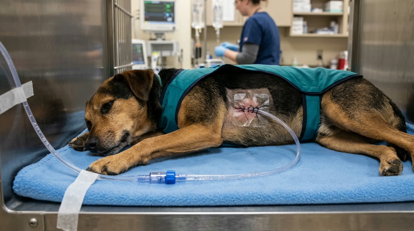
Chest Tube Placement Equipment: 16-22 Fr tube (cats); 22-32 Fr (large dogs); scalpel, hemostats, 2-0 nylon suture; Heimlich valve
Technique: 1. Sedate; bupivacaine 1 mg/kg local analgesia 2. Clip 7th-9th ICS dorsal 3. Skin incision over 10th ICS; tunnel cranially to 8th ICS 4. Puncture intercostal muscles at cranial border of rib 5. Insert tube craniodorsally; purse-string suture 6. Attach Heimlich one-way valve or suction (-5 to -10 cm H2O)
Ongoing Management
- Aspirate q4-6h or continuous suction
- Discontinue tube when air <5 mL/kg/hour for 12 hours
Pain Management
- Buprenorphine 0.02-0.03 mg/kg IV or IM q6-8h
- Fentanyl CRI 2-5 mcg/kg/hour
- Intrapleural bupivacaine 1.5 mg/kg via tube q6-8h
Surgical Management
- Spontaneous PTX recurs >50% without surgery
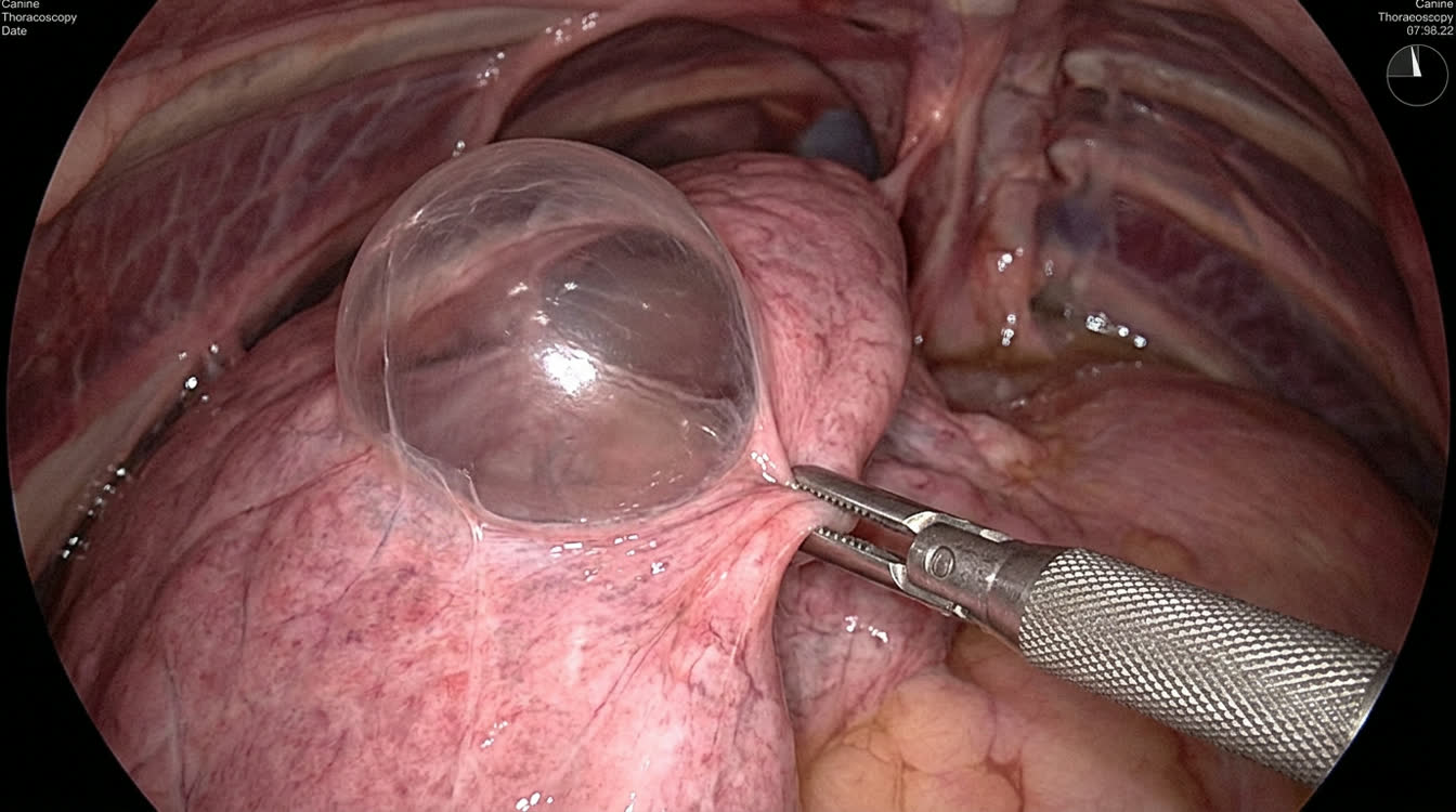
- Thoracoscopic bullae resection + partial pleurectomy
- Success rate: 70-85% long-term
Prognosis and Post-treatment Monitoring
Prognosis by Type
| Type | Prognosis | Recurrence |
|---|---|---|
| Traumatic | Good if trauma treated | <10% |
| Spontaneous -- bullae (medical) | Fair | >50% |
| Spontaneous -- bullae (surgical) | Good | 15-30% |
| Neoplasia-associated | Guarded | High |
Monitoring Post-Discharge
- Activity restriction: 2-4 weeks (traumatic), 6-8 weeks (post-surgery)
- Recheck radiographs at 1, 2, and 4 weeks
- Warning signs: increased breathing rate, restlessness, exercise intolerance
Traumatic PTX Considerations
- Concurrent injuries common: rib fractures, pulmonary contusions, hemothorax, diaphragmatic hernia
- Pulmonary contusions worsen 24-48h post-trauma; reassess radiographs at 24h
Cats with Spontaneous Pneumothorax
- Consider parasitic causes: Aelurostrongylus abstrusus, Paragonimus kellicotti
- Baermann fecal test; serology
- Treatment: fenbendazole 50 mg/kg PO q24h x 10-14 days
Related Articles:
- [Pleural Effusion in Dogs and Cats](/articles/pleural-effusion-dogs-cats)
- [Laryngeal Paralysis in Dogs](/articles/laryngeal-paralysis-dogs)
Frequently Asked Questions
How do I distinguish pneumothorax from pleural effusion on exam?
Pneumothorax: absent/hyperresonant sounds DORSALLY (air rises). Pleural effusion: absent sounds VENTRALLY (fluid sinks). Thoracic ultrasound helps: absent glide sign for pneumothorax; hyperechoic fluid for effusion.
What is tension pneumothorax and why is it dangerous?
Tension pneumothorax: one-way valve allows air to continuously enter the pleural space but not escape. Progressive pressure compresses the heart causing cardiovascular collapse. Immediately life-threatening -- decompress with needle without waiting for radiographs.
Should dogs with spontaneous pneumothorax have surgery?
Generally yes, for recurrent spontaneous pneumothorax or confirmed bullae. Conservative management has >50% recurrence rate. Thoracoscopic bullae resection with partial pleurectomy provides 70-85% long-term success.
Can a small pneumothorax be managed without a chest tube?
Small pneumothorax (<30% collapse) with mild signs can sometimes be managed with cage rest and oxygen, allowing air to resorb over 3-5 days. Any deterioration requires immediate drainage.
References
- Moores AL, et al. Outcome after surgical management of spontaneous pneumothorax in 20 dogs. Vet Surg. 2008;37(4):358-365.
- Au JJ, et al. Thoracoscopy for spontaneous pneumothorax in 12 dogs. Vet Surg. 2006;35(6):530-535.