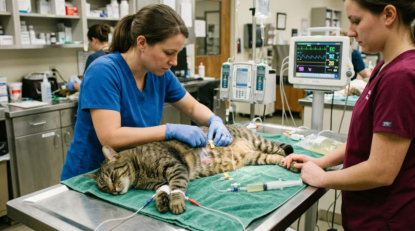
Quick Answer
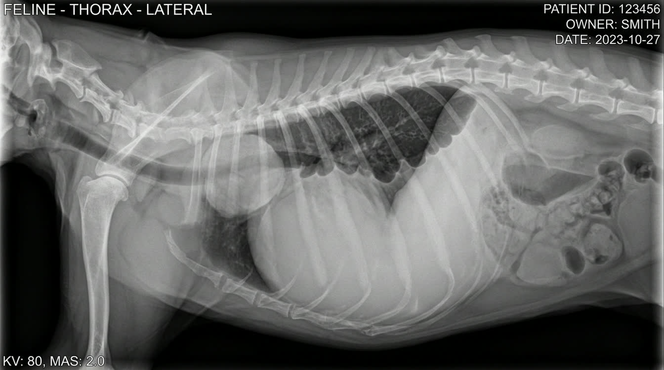
Pleural effusion is fluid accumulation in the pleural space causing respiratory distress. This guide covers species-specific causes in dogs vs cats, emergency thoracocentesis technique, cytological classification of effusion types, and management of chylothorax and pyothorax.
Key Takeaways
- ✓In cats: chylothorax, pyothorax, and FIP are most common causes
- ✓Insert needle at cranial border of the rib (7th-8th ICS) to avoid neurovascular bundle
- ✓Perform emergency thoracocentesis before full workup in dyspneic patients
- ✓Chylous effusion: pleural triglyceride > serum triglyceride confirms diagnosis
- ✓Pyothorax: chest tube lavage + 4-6 weeks culture-directed antibiotics
Causes by Species: Dogs vs Cats
Emergency Recognition Respiratory distress signs: open-mouth breathing (cats), orthopnea, extended elbows, absent ventral breath sounds, muffled heart sounds.
Causes in Dogs
| Cause | Effusion Type | Notes |
|---|---|---|
| Congestive heart failure | Modified transudate | Bilateral; elevated BNP |
| Pyothorax | Exudate | Anaerobes; Nocardia, Actinomyces |
| Chylothorax | Chylous | Mediastinal mass rule-out |
| Neoplasia | Exudate/hemorrhagic | CT for staging |
| Hypoalbuminemia | Pure transudate | Low albumin; bilateral |
Causes in Cats
| Cause | Effusion Type | Notes |
|---|---|---|
| Chylothorax | Chylous | #1 cause; idiopathic or HCM/lymphoma |
| Pyothorax | Exudate | Anaerobes; Pasteurella; bite wounds |
| FIP | Modified transudate | Yellow, fibrinous; positive Rivalta test |
| HCM | Modified transudate | Elevated proBNP |
| Mediastinal lymphoma | Mod. transudate/hemorrhagic | Young cats; anterior mediastinal mass |
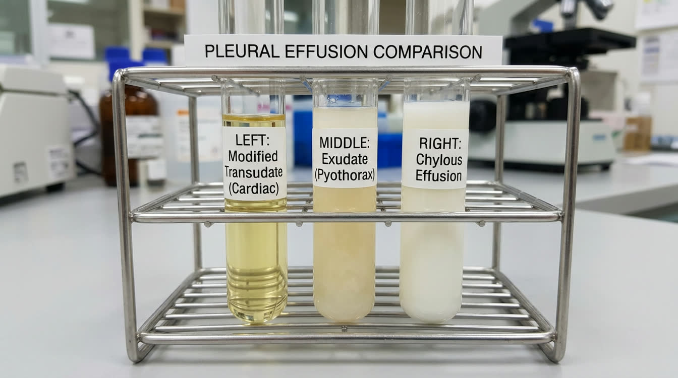
Effusion Classification
| Type | Total Protein | Cell Count | Cause |
|---|---|---|---|
| Pure transudate | <2.5 g/dL | <1000/uL | Hypoalbuminemia |
| Modified transudate | 2.5-5.0 g/dL | 1000-10000 | CHF, neoplasia |
| Exudate | >3.0 g/dL | >10000 | Infection, neoplasia |
| Chylous | Variable; TG>serum | >5000 | Lymphatic dysfunction |
| Septic exudate | >3.0 g/dL; bacteria | >50000 | Pyothorax |
Thoracocentesis Technique
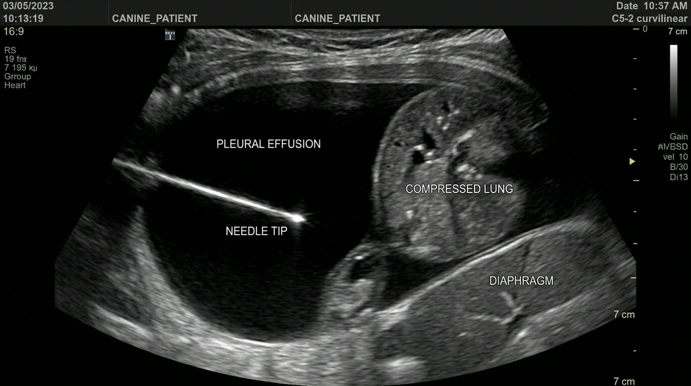
When to Perform Emergency thoracocentesis -- both diagnostic and therapeutic. Perform BEFORE full workup in respiratory distress.
Step-by-Step Technique 1. Sternal or standing recumbency 2. Site: 7th-8th ICS at costochondral junction (ventral one-third of thorax) 3. Clip and prep with chlorhexidine 4. Insert needle at the cranial border of the rib (neurovascular bundle runs caudal to each rib) 5. 19-23 gauge butterfly needle or 18-20 gauge catheter 6. Advance slowly while aspirating; drain bilaterally if needed 7. Collect EDTA tube (cytology), plain red top (chemistry/culture)
Fluid Analysis to Request
- Cell count and differential; total protein
- Culture and sensitivity (exudates)
- Triglycerides (confirm chylothorax: pleural TG > serum TG)
- Rivalta test (FIP: stir 1 drop in 0.98% acetic acid; positive = precipitates)
Chylous Effusion Management
- Initial: thoracocentesis PRN
- Rutin 50 mg/kg PO q8h: may reduce chyle production
- Low-fat diet to reduce chylomicrons
- Surgery: TDL + pericardectomy + cisterna chyli ablation; 50-80% success
Pyothorax Treatment 1. Emergency drainage; chest tube placement 2. Thoracic lavage: warm saline 10-20 mL/kg q6h through chest tube 3. IV antibiotics empirically: ampicillin-sulbactam 22 mg/kg IV q8h + enrofloxacin 5 mg/kg IV q24h 4. 4-6 weeks antibiotic therapy based on culture
Long-term Management and Prognosis
Management by Cause
| Cause | Treatment | Prognosis |
|---|---|---|
| CHF | Furosemide, pimobendan, enalapril | Fair; recurrence common |
| Chylothorax | Low-fat diet, rutin, TDL surgery | Guarded without surgery; 50-80% with TDL |
| Pyothorax | Antibiotics 4-6 weeks, lavage | Fair; 50-80% survival |
| FIP | GS-441524 antiviral | Good (>85% remission) |
| Mediastinal lymphoma | Chemotherapy | 6-12 months median |
| HCM cats | Furosemide, +/- diltiazem | Fair; recurrence common |
Post-Thoracocentesis Care
- Thoracic radiographs to confirm re-expansion
- Monitor SpO2 and respiratory rate
- Rapid re-accumulation suggests active production -- consider chest tube
Related Articles:
- [Pneumothorax in Dogs and Cats](/articles/pneumothorax-dogs-cats)
- [Laryngeal Paralysis in Dogs](/articles/laryngeal-paralysis-dogs)
Frequently Asked Questions
What are the most common causes of pleural effusion in cats vs dogs?
In cats: chylothorax (#1), pyothorax, and FIP. In dogs: CHF, pyothorax, chylothorax, and neoplasia. Species matters significantly in diagnosis and treatment planning.
Is thoracocentesis safe in a severely dyspneic patient?
Yes -- in critical patients, perform with minimal restraint in sternal recumbency with supplemental oxygen before extensive diagnostics. The procedure provides immediate relief and diagnostic information simultaneously.
How do I confirm chylous effusion?
Pleural triglyceride level higher than serum triglycerides confirms chylothorax. Visually, chylous effusion appears milky white and turbid. Cytology shows small lymphocytes and lipid vacuoles.
How much fluid can I remove at one thoracocentesis?
Remove all fluid that can be safely drained -- there is no defined limit. Monitor respiratory rate during drainage. Stop if patient becomes distressed or lung sounds return ventrally.
References
- Dempsey SM, Ewing PJ. Pathophysiology, classification, and analysis of pleural effusions. J Am Anim Hosp Assoc. 2011;47(1):1-11.
- Beatty JA, Barrs VR. Pleural effusion in the cat. J Feline Med Surg. 2010;12(9):693-707.