Quick Answer
A thorough veterinary guide to canine hip dysplasia — covering the genetic and environmental factors that contribute to the condition, how OFA and PennHIP evaluations work, and the full spectrum of treatment options including FHO, TPO, and total hip replacement surgery.
Key Takeaways
- ✓Canine hip dysplasia is a polygenic developmental condition strongly influenced by growth rate, body weight, and nutrition during the first year of life
- ✓OFA and PennHIP are complementary screening systems — PennHIP can be performed as early as 16 weeks and directly measures joint laxity
- ✓Weight management is the single most impactful intervention for both preventing and managing hip dysplasia at every stage
- ✓Surgical options include FHO (salvage), TPO (preventive in young dogs), and THR (gold standard for severe cases with over 90% success)
- ✓Consistent low-impact exercise, environmental modifications, and multimodal pain management can maintain excellent quality of life for most affected dogs
What Is Canine Hip Dysplasia?
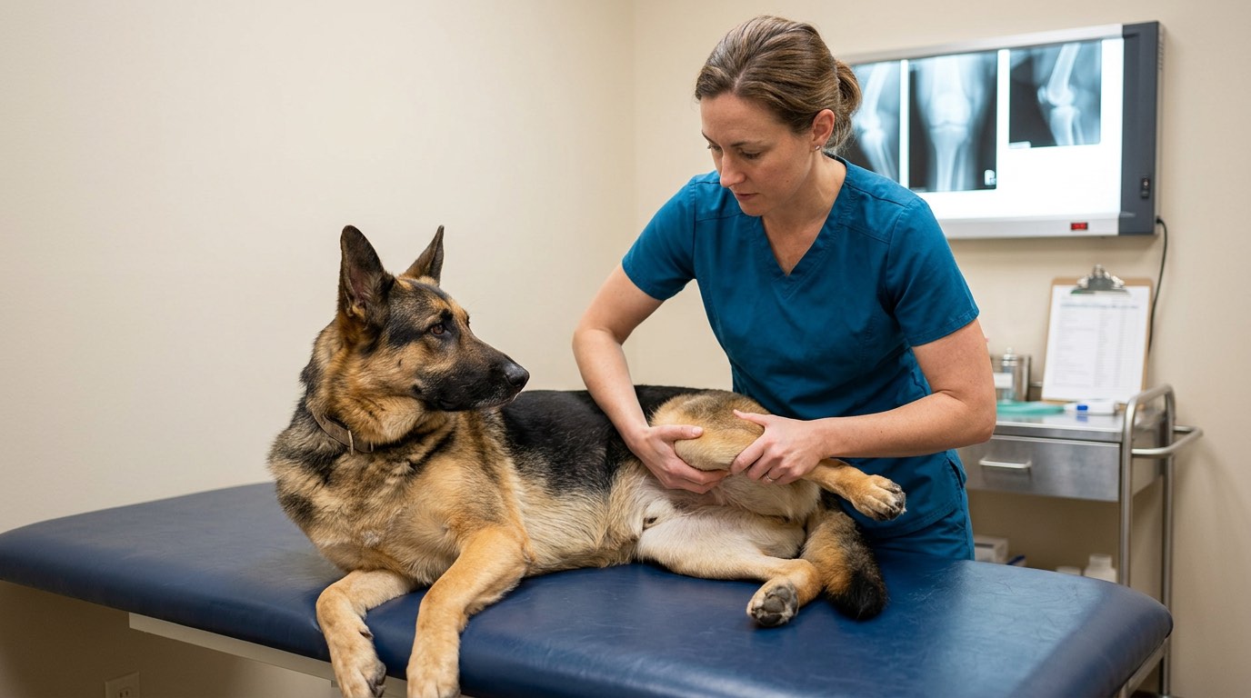
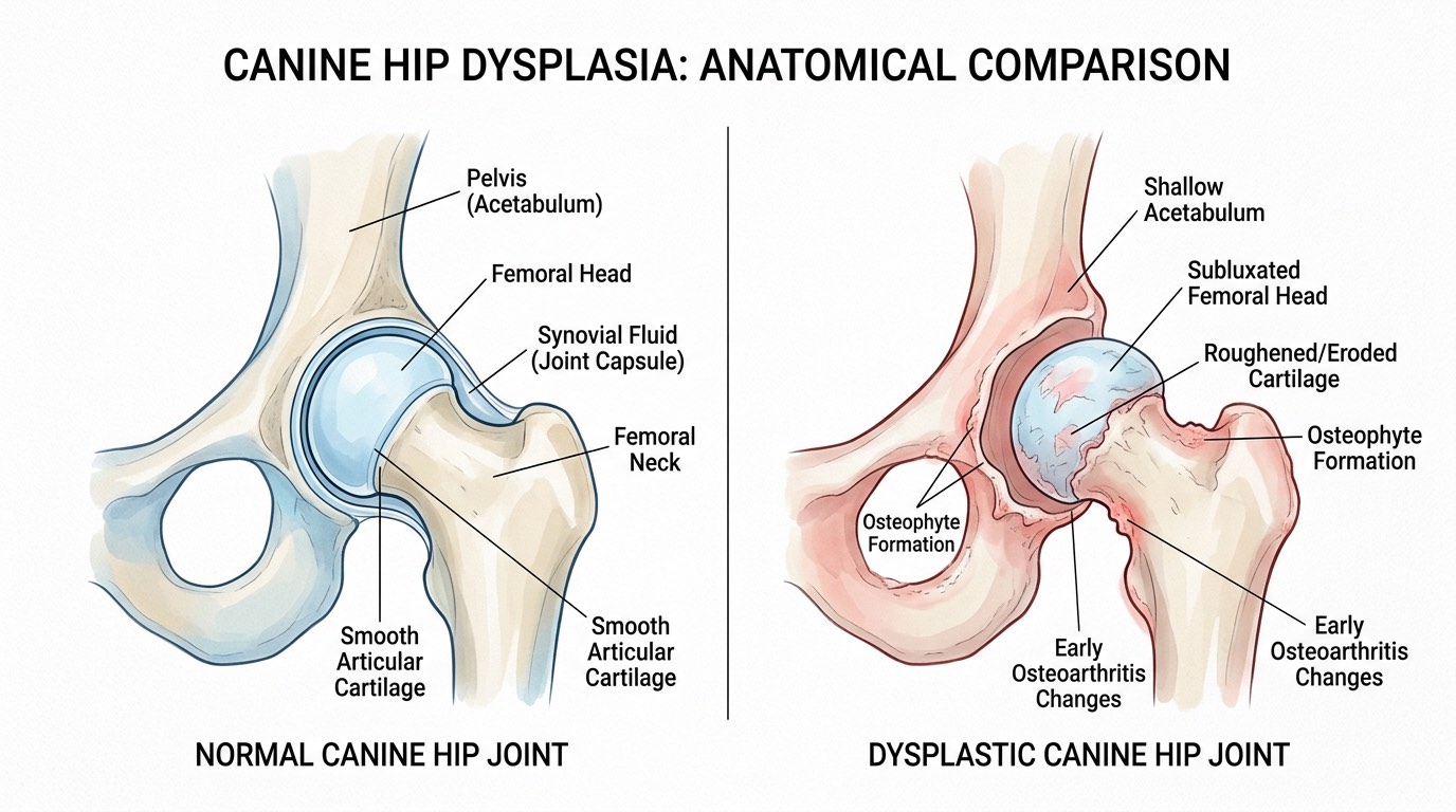
Canine hip dysplasia (CHD) is a developmental orthopedic condition in which the hip joint — a ball-and-socket joint formed by the femoral head and the acetabulum of the pelvis — fails to develop properly, resulting in joint laxity, abnormal wear, progressive cartilage degeneration, and ultimately osteoarthritis. It is one of the most common orthopedic conditions in dogs, particularly in medium to large and giant breeds, though it can occur in dogs of any size.
The condition has a strong genetic component, but its expression is influenced significantly by environmental factors including growth rate, nutrition, body weight, and exercise during the critical developmental period from puppyhood through skeletal maturity. A dog may carry the genetic predisposition for hip dysplasia yet develop clinically normal hips if environmental factors are well managed — and conversely, a genetically predisposed dog raised on a high-calorie diet with rapid growth is far more likely to develop clinical disease. Breeds with the highest prevalence include German Shepherds, Labrador Retrievers, Golden Retrievers, Rottweilers, Great Danes, Saint Bernards, and Bulldogs, though many other breeds are affected.
Clinical signs of hip dysplasia vary widely depending on the dog's age, the degree of joint laxity, and the severity of secondary osteoarthritis. Young dogs (5 to 12 months) may show reluctance to rise, a bunny-hopping gait, difficulty climbing stairs, or pain on hip extension. Older dogs with chronic dysplasia typically present with progressive hind-limb stiffness, decreased activity, muscle wasting in the hindquarters, and difficulty after rest or prolonged lying down. Some dogs with radiographic evidence of dysplasia remain clinically asymptomatic throughout life, while others with seemingly mild radiographic changes experience significant pain and functional limitation.
Genetics and Risk Factors
Hip dysplasia is a polygenic trait, meaning multiple genes contribute to a dog's susceptibility rather than a single gene that can be tested for directly. This makes selective breeding more complex — a dog with radiographically normal hips can still carry genes for dysplasia and produce affected offspring, particularly when the other parent also carries contributing genes. This is why orthopedic screening programs like OFA and PennHIP are so important for breeding stock: they provide objective, standardized assessments of hip conformation that breeders use to make informed mating decisions over multiple generations.
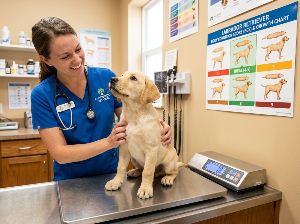
Environmental factors play a substantial modifying role in whether a genetically predisposed dog develops clinical hip dysplasia. Rapid growth during the first 3 to 10 months of life — often driven by free-choice feeding of calorie-dense puppy diets — is one of the most significant modifiable risk factors. Studies have demonstrated that controlled caloric intake during growth significantly reduces the incidence and severity of hip dysplasia in predisposed breeds. Excess body weight at any life stage increases mechanical stress on the hip joint and accelerates cartilage degradation. Inappropriate exercise during skeletal development — such as repetitive high-impact activity (jumping, stair running) on immature joints — may also contribute to joint laxity.
The interplay between genetics and environment means that responsible breeding and responsible puppy raising are both essential components of hip dysplasia prevention. Breeders should screen all breeding stock through OFA or PennHIP, select mates with consistently good hip scores across multiple generations, and provide puppy buyers with feeding and exercise guidelines. Puppy owners should feed a large-breed puppy formula with controlled calcium and calorie levels, maintain a lean body condition, and avoid excessive forced exercise until skeletal maturity (typically 12 to 18 months depending on breed).
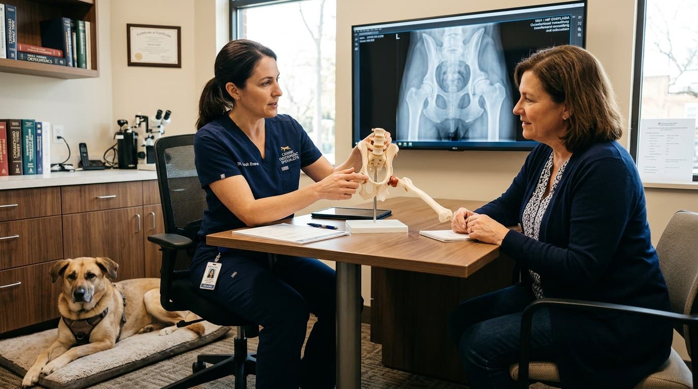
Diagnosis and Grading: OFA and PennHIP
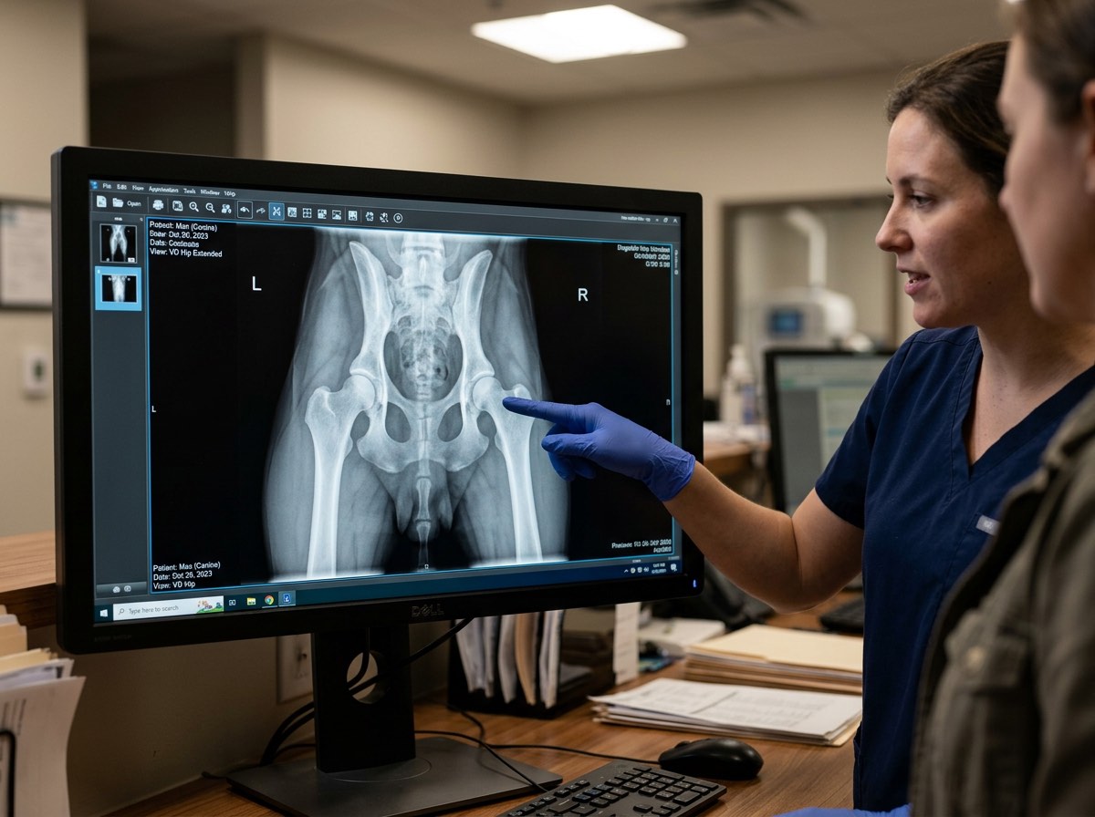
Definitive diagnosis of canine hip dysplasia requires radiographic evaluation of the hip joints, typically performed under sedation or general anesthesia to ensure proper positioning and muscle relaxation. Two primary standardized evaluation systems are used in North America: the Orthopedic Foundation for Animals (OFA) system and the University of Pennsylvania Hip Improvement Program (PennHIP).
The OFA evaluation uses a single ventrodorsal hip-extended radiograph taken at or after 24 months of age. Three independent board-certified veterinary radiologists evaluate the film and assign a consensus grade on a seven-point scale: Excellent, Good, Fair (all considered normal), Borderline, and Mild, Moderate, or Severe Dysplasia. OFA results are publicly available in a searchable database, allowing breeders and buyers to verify the hip status of dogs and their relatives. The OFA system is widely used, well-established, and accepted by most breed clubs and registries as the standard for breeding clearance. However, its primary limitation is that it evaluates joint conformation on a single static image and does not directly measure joint laxity.
PennHIP uses a distraction technique that quantifies passive hip laxity through a Distraction Index (DI), a numerical value between 0 and 1 that represents the degree to which the femoral head can be displaced from the acetabulum. A lower DI indicates a tighter hip. PennHIP can be performed as early as 16 weeks of age, providing earlier screening data. The method has been shown to be a more sensitive predictor of future osteoarthritis development than the standard OFA view. PennHIP results rank each dog against breed-specific percentile data, allowing breeders to select dogs with tighter hips relative to their breed median. Many veterinary orthopedic specialists consider PennHIP and OFA to be complementary rather than competing systems, and using both provides the most comprehensive assessment of hip joint health.
Treatment: Conservative and Surgical Options
Treatment for canine hip dysplasia spans a spectrum from conservative (non-surgical) management to advanced surgical intervention, and the appropriate approach depends on the dog's age, severity of disease, degree of pain, functional limitation, and owner considerations. Many dogs with mild to moderate hip dysplasia are managed successfully with conservative care throughout their lives, while dogs with severe joint laxity or debilitating osteoarthritis may benefit substantially from surgery.
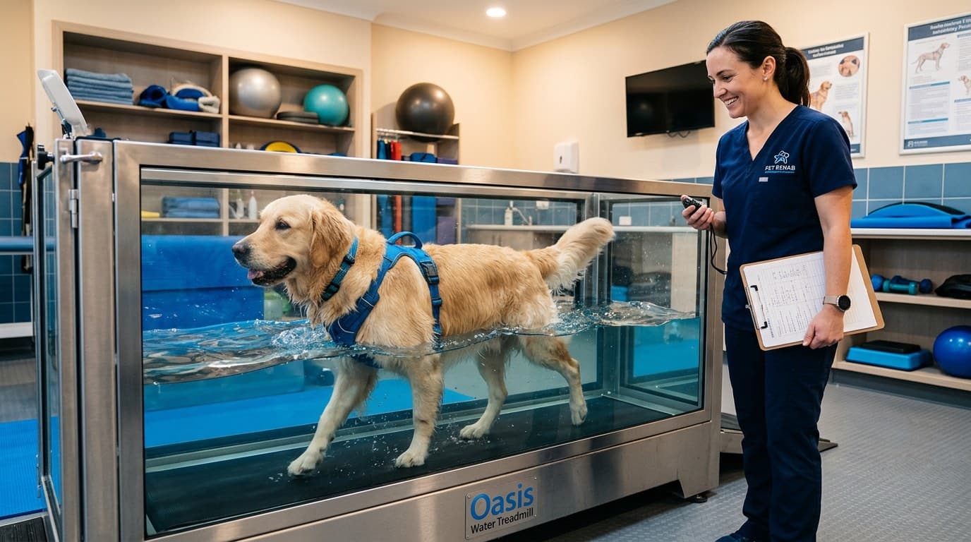
Conservative management is the foundation of hip dysplasia care for many patients. It includes weight management (maintaining a lean body condition is arguably the single most impactful intervention), controlled low-impact exercise (leash walking, swimming, underwater treadmill), non-steroidal anti-inflammatory drugs (NSAIDs such as carprofen, meloxicam, or grapiprant) for pain control, joint supplements (glucosamine, chondroitin sulfate, omega-3 fatty acids), physical rehabilitation therapy, and environmental modifications such as ramps, non-slip flooring, and orthopedic bedding. Many dogs respond well to a multimodal conservative plan, and adjustments can be made over time as the disease progresses.
Surgical options are considered when conservative management fails to provide adequate quality of life, or when the dog's age and joint status make early surgical intervention advantageous. The three primary procedures are Femoral Head and Neck Ostectomy (FHO), Triple Pelvic Osteotomy (TPO), and Total Hip Replacement (THR). FHO removes the femoral head and neck, eliminating bone-on-bone contact and allowing a fibrous pseudarthrosis (false joint) to form; it is most effective in dogs under 50 pounds and is often used as a salvage procedure. TPO is a preventive surgery performed in young dogs (typically under 10 months) with significant joint laxity but minimal arthritis — it involves cutting the pelvis in three locations and rotating the acetabular segment to improve femoral head coverage. THR replaces the entire joint with a prosthetic implant (cobalt-chrome femoral component and ultra-high-molecular-weight polyethylene acetabular cup) and is considered the gold standard for severe hip dysplasia in medium to large dogs, providing the most complete restoration of pain-free function. THR has a success rate exceeding 90% in appropriate candidates.
Recovery and Long-Term Prognosis
The prognosis for dogs with hip dysplasia varies depending on the severity of joint changes, the chosen treatment approach, and the consistency of long-term management. Dogs that undergo Total Hip Replacement typically have the best long-term outcomes, with the majority returning to normal or near-normal activity levels within three to four months of surgery. Post-operative recovery involves strict cage rest and controlled leash walks for the first 8 to 12 weeks, followed by gradual return to activity. Complications such as implant loosening, infection, and luxation of the prosthetic joint occur in fewer than 10% of cases when performed by a board-certified veterinary surgeon.
Recovery from FHO is generally good in smaller dogs but may be more variable in larger breeds. Successful FHO outcomes depend heavily on aggressive physical rehabilitation to build muscle mass around the hip, which stabilizes the pseudarthrosis and maintains limb function. Most dogs regain comfortable walking and daily activity, though some larger dogs may retain a mild gait abnormality. TPO recovery involves 8 to 12 weeks of restricted activity to allow the osteotomy sites to heal, and outcomes are best when the procedure is performed before significant arthritis develops.
For dogs managed conservatively, the condition is chronic and progressive, requiring lifelong attention to weight management, exercise modulation, pain control, and periodic veterinary reassessment. Many conservatively managed dogs live full, comfortable lives with an appropriate multimodal plan. Advances in veterinary pain management — including newer NSAID formulations, monoclonal antibody therapies (such as bedinvetmab for osteoarthritis pain), regenerative therapies, and structured rehabilitation — continue to improve quality of life for dogs living with hip dysplasia at every severity level.
What You Can Do at Home
If your dog has been diagnosed with hip dysplasia — or belongs to a breed at higher risk — there are several practical steps you can take at home to slow disease progression, reduce pain, and maximize quality of life. Weight management is the single most impactful home intervention: maintaining your dog at a lean body condition score (4 to 5 out of 9 on the standard veterinary scale) reduces mechanical stress on the hip joints by a meaningful margin. Studies by the Purina Lifespan Study demonstrated that lean-fed dogs developed osteoarthritis an average of three years later than their overfed littermates — a striking finding that underscores how powerful caloric control can be.
Provide consistent, controlled, low-impact exercise rather than sporadic bursts of high-intensity activity. Daily leash walks on flat, even surfaces are excellent. Swimming and underwater treadmill sessions (if available) provide joint-friendly conditioning that builds supporting musculature without loading the joint. Avoid activities that involve repetitive jumping, sudden direction changes, or running on hard surfaces, especially during the growth period. For older dogs with established arthritis, short, frequent walks are better tolerated than long, infrequent outings.
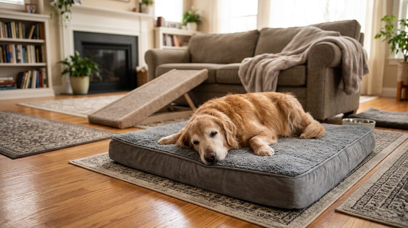
Make environmental modifications to reduce daily joint stress. Place non-slip rugs or mats on hard flooring — slipping and splaying on tile or hardwood exacerbates hip pain and can cause acute injury. Use ramps instead of stairs wherever possible, particularly for vehicle access. Provide a supportive orthopedic bed with memory foam that cushions the hips during rest. Elevate food and water bowls so your dog does not need to splay its forelimbs excessively when eating. Keep nails trimmed short, as overgrown nails alter paw placement and can shift weight-bearing patterns, increasing hip stress. Finally, administer all prescribed medications and supplements consistently, and keep up with regularly scheduled veterinary rechecks to monitor progression and adjust the management plan as needed.
Frequently Asked Questions
At what age do dogs typically show signs of hip dysplasia?
Dogs can show signs of hip dysplasia as early as 5 to 12 months of age, when rapid growth and joint laxity cause pain and gait abnormalities. However, some dogs do not show clinical signs until middle age or later, when secondary osteoarthritis becomes symptomatic. Early screening through OFA or PennHIP radiographs can detect the condition before clinical signs appear.
Can hip dysplasia be prevented through diet and exercise?
While hip dysplasia cannot be entirely prevented in genetically predisposed dogs, its severity and onset can be significantly reduced through environmental management. Feeding a controlled-calorie large-breed puppy diet, maintaining a lean body condition, and avoiding high-impact exercise during skeletal development are all proven strategies. Studies show that lean-fed dogs develop arthritis years later than overfed dogs of the same genetic background.
What is the difference between OFA and PennHIP evaluations?
OFA uses a single hip-extended radiograph taken at or after 24 months of age and assigns a subjective grade from Excellent to Severe Dysplasia. PennHIP uses a distraction technique to measure passive hip laxity as a numerical Distraction Index, and can be performed as early as 16 weeks. PennHIP is considered a more sensitive predictor of future osteoarthritis. Many specialists recommend using both systems for the most complete assessment.
Is total hip replacement worth the cost for a dog with severe hip dysplasia?
Total hip replacement is the gold standard surgical treatment for severe canine hip dysplasia, with success rates exceeding 90% when performed by a board-certified veterinary surgeon. It provides the most complete restoration of pain-free, normal function and eliminates the arthritic joint entirely. While the upfront cost is significant, many owners and veterinarians consider it the most cost-effective long-term solution for dogs with debilitating hip disease, as it often eliminates the need for ongoing medication and frequent veterinary pain management visits.
Can small dogs get hip dysplasia?
Yes, although hip dysplasia is far more common in medium, large, and giant breeds, it does occur in small-breed dogs as well. Breeds such as Pugs, French Bulldogs, and Cocker Spaniels have documented prevalence. The condition may be underdiagnosed in small dogs because their lighter body weight produces less joint stress, resulting in milder clinical signs. Any dog showing hind-limb lameness or stiffness should be evaluated radiographically regardless of size.
References
- Orthopedic Foundation for Animals. "Hip Dysplasia Statistics and Breed-Specific Data." ofa.org, 2024.
- Smith, G.K., Mayhew, P.D., Kapatkin, A.S., et al. "Evaluation of Risk Factors for Degenerative Joint Disease Associated with Hip Dysplasia in German Shepherd Dogs, Golden Retrievers, Labrador Retrievers, and Rottweilers." Journal of the American Veterinary Medical Association, 2001.
- Kealy, R.D., Lawler, D.F., Ballam, J.M., et al. "Effects of Diet Restriction on Life Span and Age-Related Changes in Dogs." Journal of the American Veterinary Medical Association, 2002.
- American College of Veterinary Surgeons. "Canine Hip Dysplasia." acvs.org, 2024.