Quick Answer
Hyperparathyroidism in dogs involves excess parathyroid hormone (PTH) causing hypercalcemia and its sequelae. This guide covers primary vs secondary hyperparathyroidism, calcium and PTH interpretation, cervical ultrasound, parathyroid surgery techniques, and post-operative hypocalcemia management.
Key Takeaways
- ✓Primary hyperparathyroidism shows elevated PTH with hypercalcemia; HHM (malignancy) shows suppressed PTH with hypercalcemia.
- ✓Cervical ultrasound identifies parathyroid adenomas >8 mm; sestamibi scintigraphy is more sensitive for smaller glands.
- ✓Post-operative hypocalcemia occurs 12-72h after parathyroid removal — start calcitriol 0.02-0.03 mcg/kg q12h pre-operatively.
- ✓Post-op monitoring: ionized calcium q4-6h for first 48h; IV calcium gluconate 5-15 mg/kg slow IV if tetany or Ca <0.8 mmol/L.
- ✓Surgical cure rate is 90-95% for solitary adenoma; treat early before renal damage becomes irreversible.
Primary vs Secondary Hyperparathyroidism: Diagnosis
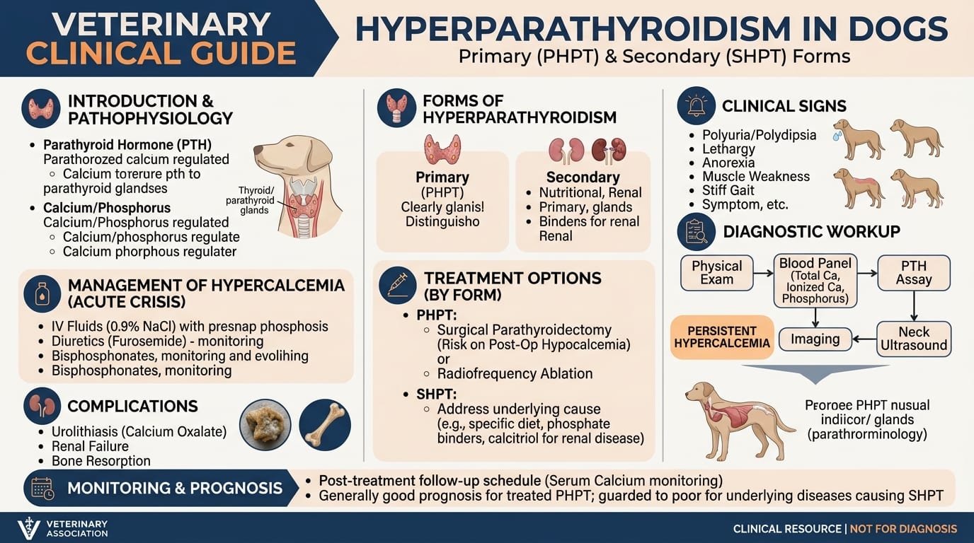
Forms of Hyperparathyroidism
| Type | PTH | Calcium | Cause |
|---|---|---|---|
| Primary HPT | High | High | Parathyroid adenoma/hyperplasia |
| Secondary (renal) HPT | High | Normal/Low | CKD — phosphate retention, calcitriol deficiency |
| Secondary (nutritional) HPT | High | Normal/Low | Low dietary calcium (raw diet) |
| Humoral hypercalcemia of malignancy (HHM) | Low/suppressed | High | Tumor PTHrP production |
| Hypercalcemia of CKD | Variable | High | Complex — calcitriol, PTH, PTHrP |
Primary Hyperparathyroidism
- Etiology: solitary parathyroid adenoma (most common, 80-90%), hyperplasia, carcinoma
- Breed predisposition: Keeshond (breed-specific hereditary form); also Golden Retriever, Labrador
- Age: typically middle-aged to older dogs (>7 years)
- Clinical signs of hypercalcemia:
Diagnostic Tests
*Serum calcium:*
- Total calcium normal: 8.9-11.4 mg/dL
- Correct total calcium: corrected Ca = Ca - albumin + 3.5
- Ionized calcium (preferred): more accurate; normal 1.12-1.40 mmol/L; hypercalcemia >1.45 mmol/L
*Parathyroid hormone (intact PTH):*
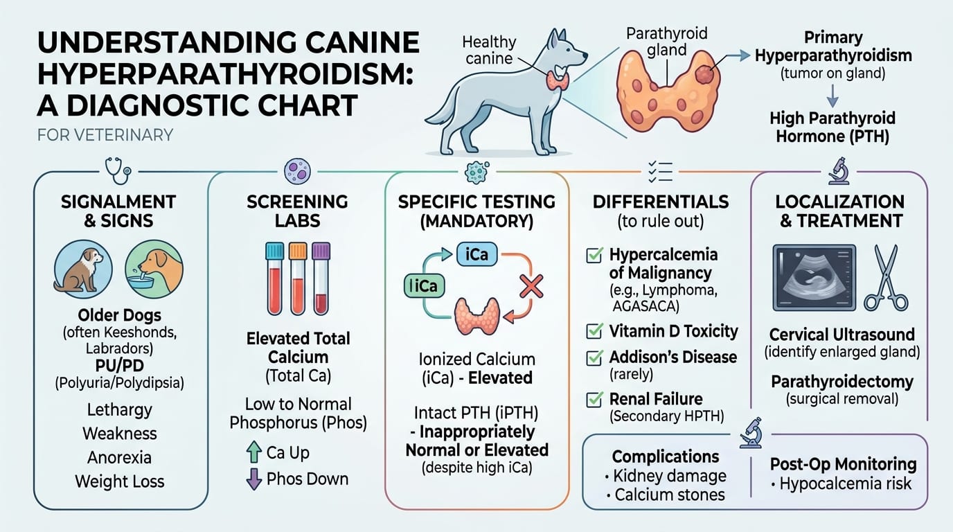
- Primary HPT: elevated or inappropriately normal in presence of hypercalcemia
- HHM (malignancy): suppressed (normal feedback response to elevated Ca)
- Secondary (renal): elevated PTH with CKD
- Test: send paired samples of ionized Ca and intact PTH to endocrine laboratory
*PTHrP (Parathyroid hormone-related protein):*
- Elevated in HHM — lymphoma, anal sac adenocarcinoma most common in dogs
- Normal in primary HPT
Causes of Hypercalcemia in Dogs — Differential Diagnosis
- C: Cancer (lymphoma, anal sac ADC, multiple myeloma)
- H: Hyperparathyroidism (primary)
- E: Excesses (vitamin D toxicosis, rodenticides, plants)
- W: Addison's disease (hypoadrenocorticism)
- T: Tertiary HPT
- O: Osteolytic bone lesion
- P: Physiologic (young growing dog — normal high end)
- M: Mycoses (blastomycosis, histoplasmosis)
- U: Unknown/Idiopathic
- D: Dehydration (artifact)
Treatment: Medical Management and Parathyroid Surgery
Medical Management of Hypercalcemia (Pre-surgical or Palliation)
For total calcium >14 mg/dL or symptomatic hypercalcemia: 1. IV 0.9% NaCl: primary treatment; saline promotes calciuresis; start 2-3x maintenance rate 2. Furosemide 1-2 mg/kg IV q4-6h: after adequate hydration; enhances calciuresis; do NOT give before rehydration 3. Prednisolone 1-2 mg/kg IV/PO q24h: if lymphoma confirmed or suspected; do NOT start before diagnosis (may mask lymphoma diagnosis on biopsy) 4. Pamidronate 1-2 mg/kg IV over 2h: bisphosphonate; inhibits osteoclast activity; used for persistent/refractory hypercalcemia; onset 2-3 days; duration 2-4 weeks 5. Salmon calcitonin 4-6 IU/kg SQ q8-12h: rapid-acting but short-lived; useful for acute severe hypercalcemia while awaiting surgery
Parathyroid Surgery — Definitive Treatment for Primary HPT Surgery is the only cure for primary hyperparathyroidism.
*Pre-operative workup:*
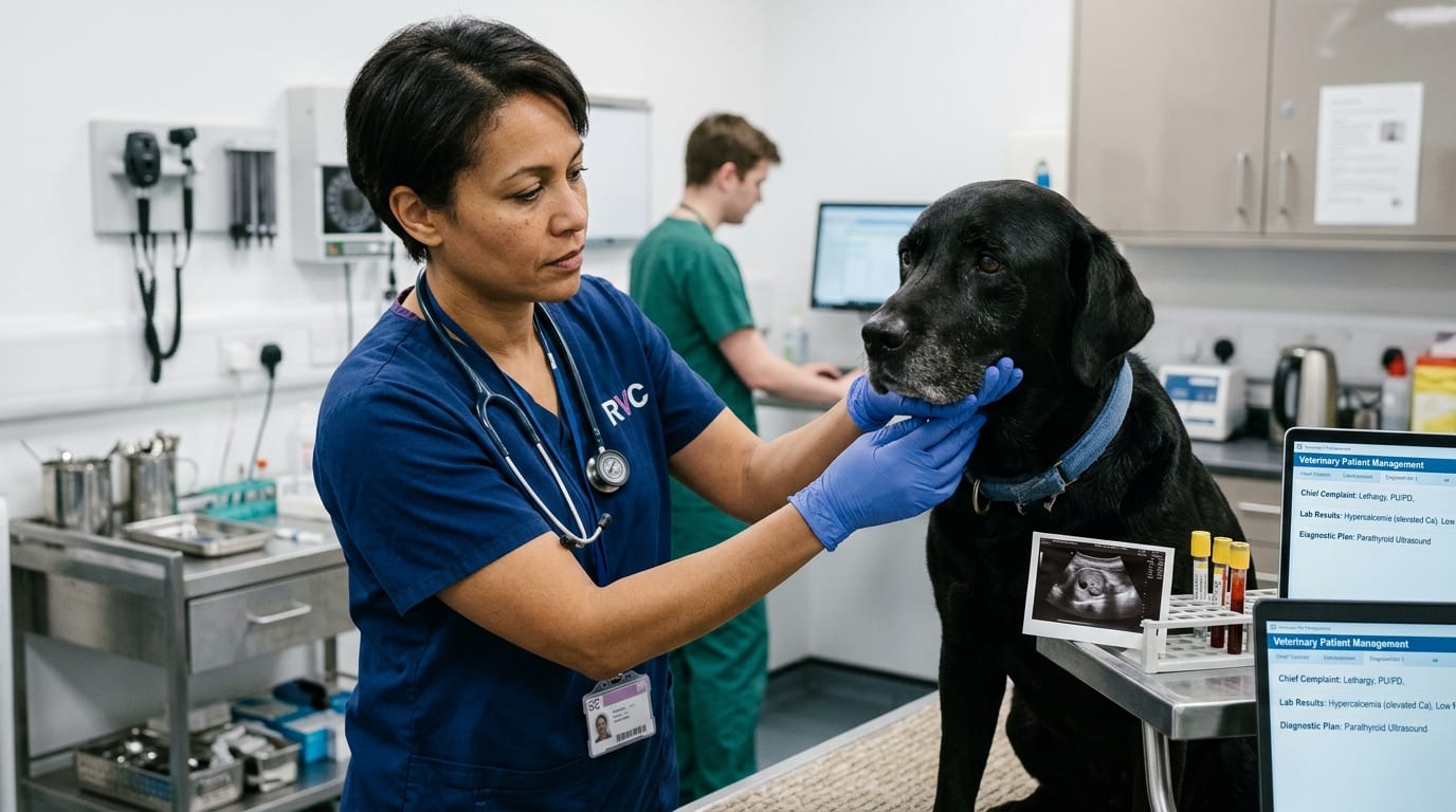
- Cervical ultrasound: identify enlarged parathyroid gland (normal <5 mm; adenoma typically 8-15 mm)
- 99m-technetium sestamibi scintigraphy: more sensitive; available at referral centers
- CBC, chemistry, ionized calcium, PTH day of surgery
- Renal function assessment (hypercalcemia-induced nephropathy)
*Surgical technique:*
- Bilateral cervical exploration (Grades I-IV approach to parathyroid glands)
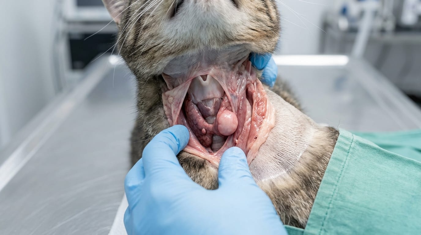
- Four parathyroid glands (two pairs): external parathyroids on thyroid surface; internal parathyroids embedded in thyroid tissue
- Identify all four glands; remove enlarged adenoma
- If no obvious adenoma: remove largest gland for pathology; remove glands showing hyperplasia
- Intraoperative PTH monitoring: rapid PTH assay 10 min post-excision — should drop >50% if all functional parathyroid tissue removed
*Ultrasound-guided percutaneous ethanol ablation (UPEA):*
- Alternative to surgery for select cases
- 99% ethanol injected directly into parathyroid adenoma under ultrasound guidance
- Success rate: ~80%; multiple sessions sometimes needed
- Preferred in high-anesthetic-risk patients
Post-operative Hypocalcemia — Life-threatening Complication After removal of a parathyroid adenoma, the remaining atrophied parathyroid glands may take days to weeks to regain function.
*Signs:* muscle tremors, facial rubbing, tetany, seizures — calcium falls rapidly post-op *Timing:* usually 12-72 hours post-surgery
*Prevention and treatment:*
- Monitor ionized calcium q4-6h for first 24-48h
- Start calcitriol (active Vitamin D3) pre-operatively: 0.02-0.03 mcg/kg PO q12h; stimulates intestinal calcium absorption; begin 1-2 days before surgery
- IV calcium supplementation if tetany or ionized Ca <0.8 mmol/L:
- Oral calcium carbonate: 25-50 mg/kg/day divided q8h; continue for weeks post-op
- Continue calcitriol until PTH recovers (may take 1-4 weeks)
Post-operative Monitoring and Long-term Outcomes
Post-operative Monitoring Schedule
| Time | Tests | Target |
|---|---|---|
| 4-6h post-op | Ionized Ca | ≥0.9 mmol/L |
| 12-24h | Ionized Ca | ≥0.9 mmol/L |
| 48-72h | Ionized Ca, PTH | Ca normalizing |
| Day 5-7 | Ionized Ca, PTH | Ca 1.0-1.2 mmol/L; PTH detectable |
| 1-2 months | Ionized Ca, total Ca, renal function | Full recovery |
Calcitriol Tapering Protocol
- Continue calcitriol 0.02-0.03 mcg/kg q12h post-op until ionized calcium consistently >1.1 mmol/L
- Reduce to q24h when Ca stable for 2 weeks
- Discontinue when ionized Ca consistently normal for 4 weeks
- Monitor ionized Ca weekly while tapering
Complications
- Recurrent laryngeal nerve damage: voice change, laryngeal paralysis (rare)
- Hypothyroidism: if thyroid tissue damaged (rare)
- Persistent hypercalcemia: incomplete removal, multi-gland disease, malignant parathyroid tumor (carcinoma)
- Renal deterioration: pre-existing hypercalcemia-induced nephropathy may not fully reverse
Prognosis
- Surgical cure rate for solitary adenoma: 90-95%
- If treated early (before significant renal damage): full recovery expected
- Uroliths (calcium oxalate): check for stone resolution/persistence on ultrasound at 3 months; dietary management post-op
- Hereditary form (Keeshond): monitor for multi-gland disease; breeding counseling recommended
Related Articles:
- [Hypercalcemia in Dogs and Cats](/articles/hypercalcemia-dogs-cats)
- [Pheochromocytoma in Dogs](/articles/pheochromocytoma-dogs)
Frequently Asked Questions
What is the difference between primary and secondary hyperparathyroidism in dogs?
Primary hyperparathyroidism means a parathyroid gland is overproducing PTH autonomously (usually an adenoma). This causes hypercalcemia and suppressed calcium-regulation feedback. Secondary hyperparathyroidism means the parathyroids are producing excess PTH as a compensatory response — usually to chronic kidney disease (reduced phosphate excretion and calcitriol production), which drives PTH upward. The key diagnostic difference: primary HPT shows elevated PTH with hypercalcemia; secondary HPT shows elevated PTH with normal or low calcium.
What happens if post-operative hypocalcemia is not treated?
Untreated post-operative hypocalcemia (ionized calcium <0.8 mmol/L) can cause life-threatening muscle tetany, laryngospasm, and seizures within 12-72 hours of parathyroid surgery. This is why calcitriol should be started 1-2 days before surgery, and ionized calcium must be monitored every 4-6 hours in the immediate post-operative period. IV calcium gluconate should always be available in the post-surgical patient.
Can hypercalcemia damage the kidneys?
Yes — sustained hypercalcemia causes calcium deposition in renal tubular cells (nephrocalcinosis), reduces renal blood flow, and impairs urine concentrating ability. Chronic hypercalcemia can cause irreversible CKD. This is why treating primary hyperparathyroidism promptly, before renal function deteriorates, improves long-term outcomes significantly.
Is surgery always required for primary hyperparathyroidism in dogs?
Surgery or ultrasound-guided percutaneous ethanol ablation (UPEA) is the only cure. Medical management with IV fluids and bisphosphonates can stabilize hypercalcemia temporarily but does not remove the autonomous PTH-producing gland. For dogs that are poor surgical candidates, UPEA offers an 80% success rate alternative. Long-term medical palliation without definitive treatment leads to progressive renal damage.
References
- Feldman EC, Nelson RW. Canine and Feline Endocrinology and Reproduction. 3rd ed. Saunders; 2004.
- Gear RN, et al. Primary hyperparathyroidism in 29 dogs. J Small Anim Pract. 2005;46(1):8-16.
- Sueda MT, Stefanacci JD. Ultrasound evaluation of the parathyroid glands in two hypercalcemic dogs. Vet Radiol Ultrasound. 2000;41(5):448-452.