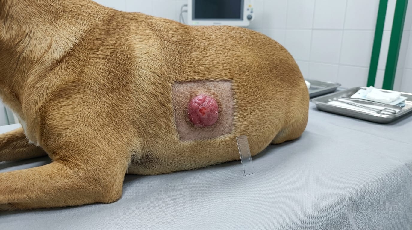
Quick Answer
Mast cell tumors (MCT) are the most common malignant skin tumor in dogs. This guide covers histological grading systems (Patnaik and Kiupel), surgical planning with adequate margins, medical management with Palladia (toceranib) and prednisone, c-KIT mutation testing, and regional lymph node staging.
Key Takeaways
- ✓Kiupel 2-grade system (low vs high) provides better reproducibility than 3-grade Patnaik
- ✓Always aspirate regional lymph nodes -- Stage II (node-positive) changes treatment to systemic therapy
- ✓Surgical margins: 2-3 cm lateral + one fascial plane deep for most MCT
- ✓Palladia 2.75 mg/kg EOD: c-KIT exon 11 mutations predict best response (~80%)
- ✓Premedicate with diphenhydramine + famotidine before MCT surgery
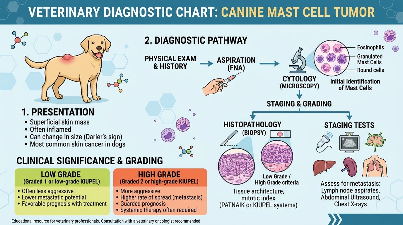
Histological Grading and Staging
Overview MCT represent 16-21% of all canine skin tumors. Biological behavior ranges from benign Grade I to highly malignant Grade III. Always perform FNA before surgery -- sensitivity >90%.
Patnaik Grading (3 Grades -- Traditional)
| Grade | Metastatic Rate | Median Survival |
|---|---|---|
| Grade I (well-differentiated) | <5% | >1500 days |
| Grade II (moderate) | 5-20% | ~500 days (variable) |
| Grade III (poorly differentiated) | >75% | ~100-200 days |
Kiupel Grading (2 Grades -- Increasingly Preferred)
| Grade | Criteria | Behavior |
|---|---|---|
| Low grade | <7 mitoses/10 HPF; no multinucleated cells; no karyomegaly | Low metastatic risk |
| High grade | >=7 mitoses/10 HPF OR >3 multinucleated cells OR karyomegaly | High metastatic risk; systemic therapy |
Staging
| Stage | Definition |
|---|---|
| I | Single tumor, no node involvement |
| II | Node involvement |
| III | Multiple tumors or large infiltrative |
| IV | Distant metastases |
Lymph Node Evaluation -- Critical
- Always aspirate regional lymph nodes before or at surgery
- Node-positive (Stage II): systemic therapy warranted
- Abdominal ultrasound for spleen/liver in high-grade tumors
c-KIT Mutation Testing
- c-KIT mutations in 15-40% of MCT
- Exon 11 mutations: predict ~80% response to Palladia
- Request IHC for c-KIT and PCR mutation testing on histopath sample
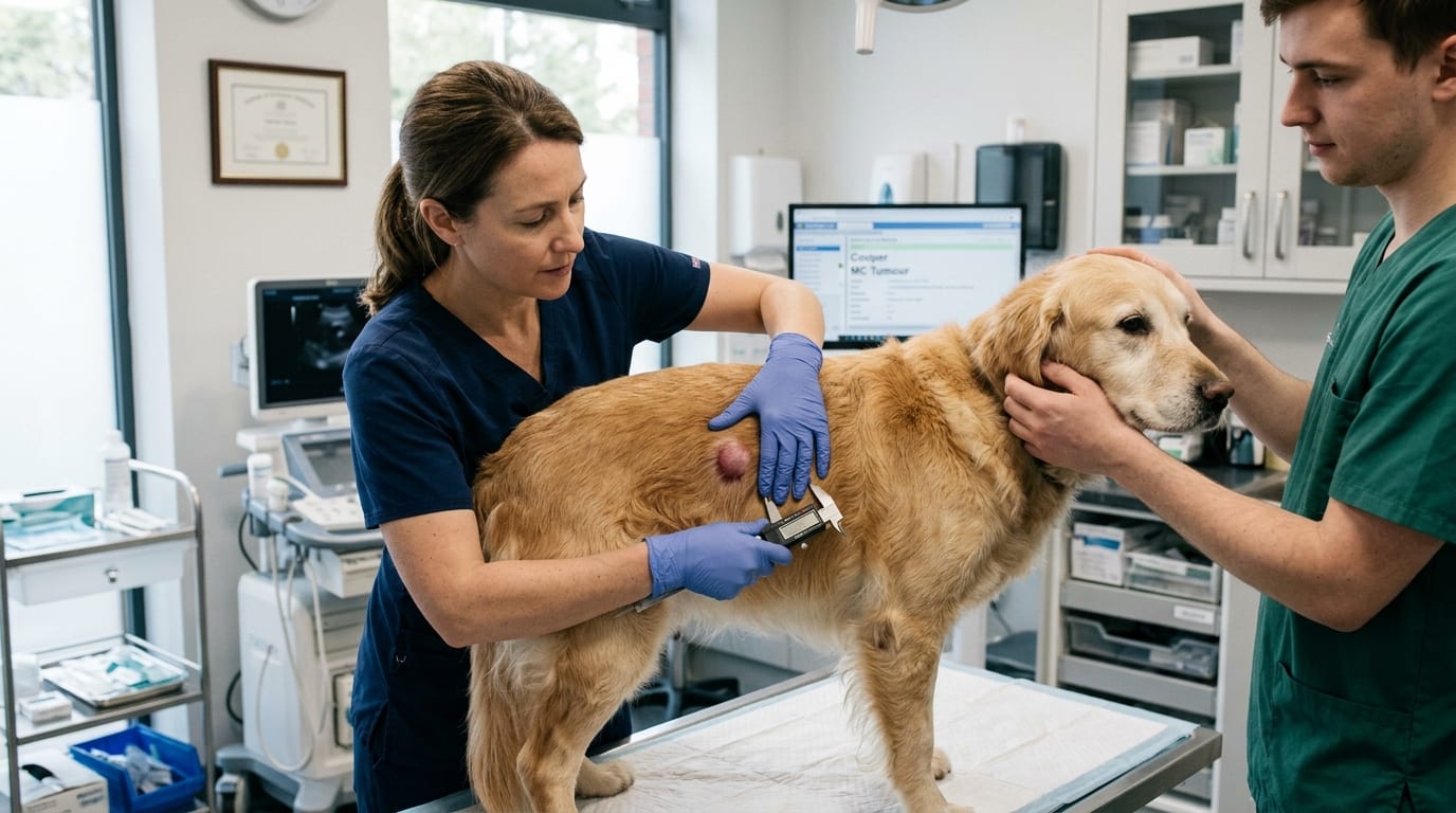
Surgical Management: Margins and Incomplete Excision
Surgical Margins
| Grade | Lateral Margin | Deep Margin |
|---|---|---|
| Low grade (Kiupel) / Grade I | 1-2 cm | One fascial plane |
| Grade II / unknown grade | 2-3 cm | One fascial plane |
| High grade (Kiupel) / Grade III | 3+ cm | Two fascial planes |
Incomplete Excision Options 1. Re-excision (best if feasible): 3 cm lateral + deeper plane 2. Radiation therapy: effective for positive margins 3. Palladia +/- prednisone: if re-excision not feasible 4. Active surveillance: low-grade narrow margins, recheck q4-8 weeks
Preoperative Preparation
- Diphenhydramine 2 mg/kg IM 30 min before (H1 blocker)
- Famotidine 0.5 mg/kg IV (H2 blocker)
- Dexamethasone SP 0.1 mg/kg IV (reduces swelling and histamine effects)
- Avoid aggressive tumor manipulation (degranulation risk)
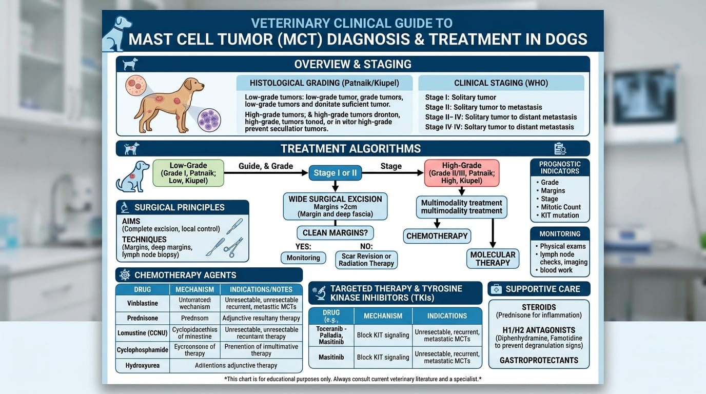
Medical Therapy: Palladia, Prednisone, and Vinblastine
Prednisone
- Adjuvant/metastatic: 1-2 mg/kg PO q24h; taper to 0.5 mg/kg q48h after 4-8 weeks
- Inoperable/palliative: 2 mg/kg PO q24h
Palladia (Toceranib Phosphate) -- FDA-Approved
| Parameter | Details |
|---|---|
| Dose | 2.75 mg/kg PO every other day (MWF) |
| Response with c-KIT exon 11 mutation | ~80% |
| Response without mutation | ~30-40% |
| Overall objective response | 40-60% |
| Side effects | GI, neutropenia, hypoalbuminemia, protein-losing nephropathy, limb edema |
| Monitoring | CBC, chemistry, UPC q2-4 weeks |
Additional Options
- Vinblastine 2 mg/m2 IV weekly +/- prednisone: adjuvant for high-grade; 40-50% response
- Lomustine (CCNU) 60-70 mg/m2 PO q3-6 weeks: Grade III or metastatic MCT
Histamine Management
- Diphenhydramine 2 mg/kg PO q8-12h (H1 blocker)
- Famotidine 0.5-1 mg/kg PO q12-24h or omeprazole 0.5-1 mg/kg PO q24h (GI protection)
- Sucralfate 0.5-1 g/dog PO q8h with food: if GI ulceration suspected
Related Articles:
- [Lymphoma Treatment in Dogs](/articles/lymphoma-dogs-treatment)
- [Spironolactone in Dogs and Cats](/articles/spironolactone-dogs-cats)
Frequently Asked Questions
What is the difference between Patnaik and Kiupel grading?
Patnaik uses three grades (I, II, III). Grade II historically caused inconsistency because it encompasses a wide behavior range. The Kiupel 2-grade system (low vs high grade) uses specific objective criteria -- primarily mitotic count, multinucleated cells, and karyomegaly -- providing better reproducibility. Most pathologists now report both systems.
How important is lymph node aspiration?
Critical -- lymph node involvement indicates Stage II disease and typically warrants systemic therapy (Palladia or vinblastine/prednisone). Always aspirate the regional draining lymph node before or at surgery, even if it appears normal.
When should Palladia be used?
For high-grade or Stage II/III disease, incomplete excision where re-excision is not possible, recurrent disease, or systemic/metastatic disease. c-KIT exon 11 mutations predict best response (~80%), but even without mutations, ~40% objective response rate is seen.
Do I need to premedicate before MCT surgery?
Yes -- diphenhydramine 2 mg/kg IM + famotidine 0.5 mg/kg IV given 30 minutes before surgery. Dexamethasone 0.1 mg/kg IV also helps. Avoid aggressive tumor manipulation intraoperatively to minimize degranulation.
References
- Kiupel M, et al. Proposal of a 2-tier grading system for canine cutaneous mast cell tumors. Vet Pathol. 2011;48(1):147-155.
- London CA, et al. Prospective evaluation of toceranib phosphate in dogs with mast cell tumors. J Vet Intern Med. 2009;23(1):107-117.